

A Comprehensive Guide: Vasectomy vs Orchiectomy Orchiectomy is a surgical procedure designed for permanent sterilization in male dogs, intended to prevent reproduction. While it’s a routine procedure, it’s often misunderstood or surrounded by misconceptions. In this comprehensive guide, we’ll delve into the intricacies of orchiectomy, highlighting its differences from vasectomy, and exploring its relationship with canine health, particularly concerning conditions like testicular cancer and prostatic disease. Understanding Orchiectomy: Orchiectomy involves the removal of the testicles, the primary organs responsible for producing sperm and testosterone in male dogs. By eliminating these organs, the dog becomes sterile, unable to reproduce. Orchiectomy is typically performed by a veterinarian under general anesthesia and is considered a safe and effective way to control pet population and behavior. Compared to alternative methods like hormone injections or contraceptives, orchiectomy provides a permanent solution with various health benefits. Differences Between Orchiectomy and Vasectomy: While both procedures involve altering the reproductive system of male dogs, they serve distinct purposes. Orchiectomy specifically refers to the removal of the testicles, whereas vasectomy involves blocking or cutting the vas deferens, the tubes that carry sperm from the testicles to the urethra. Orchiectomy results in permanent infertility, while vasectomy allows for the continued production of sperm without fertilization. Orchiectomy is primarily aimed at preventing unwanted litters and addressing behavioral issues, while vasectomy may be performed for medical reasons or as a reversible form of contraception. The Relationship Between Canine Health and Hormonal Balance: Hormonal balance plays a crucial role in maintaining overall health and well-being in dogs. Testosterone, the primary male sex hormone produced by the testicles, influences various physiological processes, including reproductive function, behavior, and musculoskeletal development. While testosterone is essential for normal growth and development, imbalances or excess levels may contribute to certain health conditions in male dogs. Testicular Cancer: Testicular cancer is relatively common in unneutered male dogs, with certain breeds being predisposed to this condition. Orchiectomy significantly reduces the risk of testicular tumors by removing the organs where these tumors originate. While the exact causes of testicular cancer are not fully understood, hormonal factors, genetic predisposition, and environmental influences may play a role in its development. Prostatic Disease: The prostate gland, located near the base of the bladder, is susceptible to various conditions, including prostatic enlargement and inflammation. Testosterone influences the growth and function of the prostate, with elevated levels potentially contributing to prostatic disease. Orchiectomy reduces testosterone levels, which may help prevent conditions like benign prostatic hyperplasia (BPH) or prostatitis, inflammation of the prostate gland. Conclusion: Orchiectomy is a safe and effective way to control pet population, address behavioral issues, and promote overall canine health and well-being. While it differs from vasectomy in its scope and implications, both procedures highlight the importance of responsible pet ownership and veterinary care. Additionally, hormonal balance plays a significant role in maintaining canine health, with orchiectomy offering various benefits, including reducing the risk of testicular cancer and prostatic disease. Any other doubt? We are here to help, Contact us now. Contact Us Preventive Medicine Click here Pet Surgery Click here Alternative Medicine Click here Pet Dentistry Click here

Lyme Disease in Dogs
Lyme Disease in Dogs: Understanding, Preventing, and Treating Lyme disease, caused by the bacterium Borrelia burgdorferi, is a prevalent tick-borne illness affecting both humans and canines. In dogs, this disease manifests with symptoms ranging from mild discomfort to severe complications if left untreated. Understanding Lyme disease, its prevention, and treatment is crucial for every dog owner. Understanding Lyme Disease in dogs: Lyme disease is primarily transmitted through the bite of infected black-legged ticks, also known as deer ticks. These ticks are commonly found in grassy, wooded areas, and can lat onto dogs during outdoor activities like hiking or playing in the yard. Once infected, the bacteria can spread through the dog’s bloodstream, leading to various symptoms. Lyme disease symptoms in dogs can vary widely, making diagnosing it challenging. Common signs include lameness, joint pain, fever, lethargy, loss of appetite, and swollen lymph nodes. In some cases, dogs may develop kidney complications or neurological issues if the disease progresses untreated. Any other doubt? We are here to help, Contact us now. Contact Us Prevention Measures: Preventing Lyme disease starts with reducing exposure to ticks. Here are some effective prevention measures: Tick Control: Use tick control products recommended by veterinarians, including topical treatments, collars, or oral medications. These products can kill ticks on contact or repel them, reducing the risk of transmission. Regular Tick Checks: After outdoor activities, thoroughly check your dog for ticks, paying close attention to areas like ears, armpits, and between toes. Promptly remove any ticks using fine-tipped tweezers, grasping the tick close to the skin and pulling straight out with steady pressure. Environmental Management: Keep your yard well-maintained by regularly mowing grass, removing leaf litter, and creating barriers between wooded areas and play areas. Consider using tick control products in outdoor spaces frequented by your dog. Vaccination: Talk to your veterinarian about the Lyme disease vaccine for dogs. While it may not offer complete protection, vaccination can help reduce the severity of symptoms if your dog is exposed to the bacteria. Education: Learn about the local prevalence of Lyme disease and the peak times for tick activity in your area. This knowledge can help you adjust outdoor activities and prevention strategies accordingly. Treatment for Lyme Disease for dogs: If your dog is diagnosed with Lyme disease, early treatment is essential to prevent complications. Treatment typically involves antibiotics to eliminate the bacteria from the dog’s system. Commonly prescribed antibiotics include doxycycline or amoxicillin, administered over several weeks. In addition to antibiotics, supportive care may be necessary to manage symptoms such as pain and inflammation. Your veterinarian may recommend anti-inflammatory medications, joint supplements, and rest to help your dog recover comfortably. Regular follow-up appointments with your veterinarian are crucial to monitor your dog’s progress and ensure the effectiveness of treatment. In some cases, dogs may require additional testing to assess kidney function or other potential complications associated with Lyme disease. In conclusion, Lyme disease poses a significant health risk to dogs. Still, with proper understanding, prevention measures, and prompt treatment, you can help protect your furry companion from this potentially debilitating illness. Stay vigilant, stay informed, and prioritize your dog’s health and well- being in every outdoor adventure. By taking proactive steps to prevent tick bites and seeking veterinary care at the earliest signs of Lyme disease, you can ensure your dog leads a happy, healthy life free from this preventable illness. Related Services Preventive Medicine Click here Pet Surgery Click here Alternative Medicine Click here Pet Dentistry Click here
Feline Leukemia and Feline Immunodeficiency Virus
Understanding Feline Leukemia and Feline Immunodeficiency Virus (FIV): Vaccination Protocols and Management Feline leukemia virus (FeLV) and feline immunodeficiency virus (FIV) are two significant viral infections that affect cats worldwide. These diseases weaken the immune system, making affected cats susceptible to various infections and health complications. Understanding the nature of FeLV and FIV, along with proper vaccination protocols and management strategies, is crucial for cat owners and veterinarians alike. Feline Leukemia Virus (FeLV): FeLV is a retrovirus that infects cats and can lead to various health issues, including anemia, immune suppression, and the development of certain cancers, such as lymphoma and leukemia. The virus is primarily transmitted through close contact with infected cats, particularly through mutual grooming, sharing food and water bowls, and mating. Symptoms of FeLV infection can vary widely and may include lethargy, weight loss, poor coat condition, fever, and recurrent infections. However, some cats may not show symptoms for months or even years after infection. FeLV-positive cats are at an increased risk of developing severe illnesses, and the prognosis for affected cats depends on various factors, including their overall health and the presence of concurrent diseases. Feline Immunodeficiency Virus (FIV): FIV is another retrovirus that affects cats, leading to progressive immune system dysfunction. Unlike FeLV, which can cause severe illness relatively quickly, FIV often progresses slowly over several years. The virus is primarily transmitted through bite wounds inflicted during aggressive encounters between cats, particularly among intact males engaged in territorial disputes. Symptoms of FIV infection can include recurrent infections, gingivitis, weight loss, and a decline in overall health. As with FeLV, some cats may remain asymptomatic for extended periods after infection. While there is no cure for FIV, supportive care and management can help affected cats live relatively comfortable lives for an extended period. Any other doubt? We are here to help, Contact us now. Contact Us Vaccination Protocols: Vaccination plays a crucial role in preventing FeLV and FIV infections in cats. Here is a recommended vaccination protocol for both diseases: FeLV Vaccination: Kittens should receive their first FeLV vaccination between 8 to 9 weeks of age, followed by a booster vaccine three to four weeks later. Cats at high risk of exposure, such as those living in multi-cat households or allowed outdoor access, should receive annual booster vaccinations thereafter. Indoor-only cats with minimal exposure to other cats may require less frequent vaccination, depending on their risk factors. FIV Vaccination: The FIV vaccine is less commonly used than the FeLV vaccine due to several factors, including limited efficacy and potential interference with FIV testing. However, some veterinarians may recommend the FIV vaccine for cats at high risk of exposure, such as outdoor cats with a history of fighting. The vaccination protocol typically involves an initial series of two doses given three to four weeks apart, followed by annual booster vaccinations. It’s essential to consult with your veterinarian to determine the most appropriate vaccination protocol for your cat based on their lifestyle, risk factors, and overall health status. Management Strategies: In addition to vaccination, effective management strategies can help reduce the risk of FeLV and FIV transmission and improve the overall health and well-being of affected cats: Testing: Testing for FeLV and FIV is recommended for all cats, particularly those with unknown vaccination status or potential exposure to infected individuals. Testing should be performed before initiating vaccination protocols and whenever new cats are introduced into multi-cat households. Indoor Housing: Keeping cats indoors can significantly reduce their risk of exposure to FeLV and FIV, particularly if they live in areas where infected cats are prevalent or engage in territorial behavior. Spaying and Neutering: Sterilizing cats can reduce the likelihood of fighting and territorial disputes, decreasing their risk of FIV transmission through bite wounds. Regular Veterinary Care: Routine veterinary examinations, preventive care, and parasite control are essential for maintaining the health of all cats, particularly those at increased risk of FeLV and FIV infection. In conclusion, understanding the nature of feline leukemia virus (FeLV) and feline immunodeficiency virus (FIV), along with implementing appropriate vaccination protocols and management strategies, is crucial for safeguarding the health of cats. Cat owners can help protect their feline companions from these potentially devastating diseases by working closely with veterinarians and adopting preventive measures. Remember, early detection, vaccination, and proper management are key to ensuring cats lead long, healthy lives free from the threat of FeLV and FIV. Related Services for cats Preventive Medicine Click here Pet Surgery Click here Alternative Medicine Click here Pet Dentistry Click here

Dry eye
Understanding Dry Eye Syndrome (DES) in Brachycephalic Breeds What is DES? Dry Eye Syndrome occurs when there’s insufficient tear production or poor tear quality, resulting in dryness and inflammation of the eyes. In brachycephalic breeds, the anatomical structure, particularly the shortened nasal passages and shallow eye sockets, contributes to the development of DES. These structural abnormalities can lead to improper tear drainage and reduced blinking, exacerbating the risk of tear film instability and subsequent dryness of the cornea and conjunctiva. Symptoms of Dry Eye Syndrome: Owners of brachycephalic pets should be vigilant for signs of Dry Eye Syndrome, which may include: Excessive blinking or squinting: Brachycephalic breeds may exhibit frequent blinking or squinting as a response to ocular discomfort caused by dryness. Redness and inflammation: Irritation of the eyes due to dryness can manifest as redness and inflammation of the conjunctiva (the membrane covering the inner eyelids) and the cornea. Thick, mucoid discharge: DES can lead to the production of thick, stringy discharge from the eyes, indicating a lack of adequate tear lubrication. Corneal ulcers or erosions: Without proper tear film protection, the cornea becomes vulnerable to damage, potentially resulting in ulcers or erosions, which may lead to pain and vision impairment. Treatment Options for Dry Eye Syndrome: Managing Dry Eye Syndrome in brachycephalic breeds requires a comprehensive approach aimed at promoting tear production, reducing inflammation, and protecting the ocular surface. Treatment options may include: Artificial tear supplements: Lubricating eye drops or ointments can help replace the deficient tear film and alleviate dryness and discomfort. These supplements may need to be administered multiple times a day, especially in severe cases. Topical medications: Anti-inflammatory eye drops or ointments containing immunosuppressive agents like cyclosporine or tacrolimus may be prescribed to reduce inflammation and promote tear production. Punctal occlusion: In some cases, a veterinary ophthalmologist may recommend punctal occlusion, a procedure to block the tear drainage ducts temporarily or permanently, thereby increasing the retention of natural tears on the ocular surface. Systemic medications: Oral medications such as antibiotics or immunosuppressive drugs may be prescribed to address underlying causes or complications associated with DES, such as secondary bacterial infections or immune-mediated inflammation. Surgical interventions: In severe cases of corneal damage or persistent dryness unresponsive to medical management, surgical procedures such as conjunctival grafting or keratoplasty (corneal transplantation) may be considered to restore ocular surface integrity and function. Importance of Regular Veterinary Check-Ups: Regular veterinary check-ups are essential for brachycephalic breeds affected by Dry Eye Syndrome for several reasons: Early detection: Routine eye examinations by a veterinarian can help identify signs of DES at an early stage, allowing for prompt intervention and better treatment outcomes. Monitoring progression: DES is a chronic condition that requires ongoing management. Regular check-ups enable veterinarians to monitor the progression of the disease, adjust treatment protocols as needed, and address any emerging complications. Prevention of complications: Left untreated, Dry Eye Syndrome can lead to serious complications such as corneal ulcers, scarring, and vision loss. Regular veterinary care can help prevent or minimize these complications through timely intervention and proactive management. Educational opportunities: Veterinary visits provide an opportunity for pet owners to learn about the importance of proper eye care, including strategies for administering medications, recognizing signs of ocular discomfort, and maintaining ocular hygiene. Any other doubt? We are here to help, Contact us now. Contact Us The Chronicity of Dry Eye Syndrome It’s important for pet owners to understand that Dry Eye Syndrome is a chronic condition that requires lifelong management. Even with appropriate treatment, relapses or flare-ups may occur, necessitating ongoing monitoring and adjustments to the treatment plan. Additionally, brachycephalic breeds may be predisposed to other ocular issues, such as corneal ulcers, entropion (inward rolling of the eyelids), or distichiasis (abnormal eyelash growth), which can further complicate the management of DES. In conclusion, Dry Eye Syndrome poses a significant health concern for brachycephalic breeds of dogs and cats, highlighting the importance of proactive veterinary care and diligent management. By working closely with veterinarians, pet owners can help ensure the optimal ocular health and well-being of their beloved brachycephalic companions, minimizing discomfort, preserving vision, and enhancing quality of life. Related Services for Pets Preventive Medicine Click here Pet Surgery Click here Alternative Medicine Click here Pet Dentistry Click here

Leptospirosis
Leptospirosis What is Leptospirosis? Leptospirosis, often abbreviated as “lepto,” is a bacterial infection caused by various strains of the Leptospira bacteria. While it can affect humans and a variety of animals, including rodents, livestock, and wildlife, dogs are particularly susceptible. This zoonotic disease poses significant health risks to both dogs and humans, making it essential for dog owners to understand the importance of vaccination in preventing its spread. The Leptospira bacteria thrive in warm, moist environments, such as standing water, mud, and soil contaminated with the urine of infected animals. Dogs can become infected through direct contact with contaminated water or soil, ingestion of infected tissues, or exposure to the urine of infected animals, including rodents and wildlife. Additionally, the bacteria can enter the body through mucous membranes, such as the eyes, nose, or mouth, or through breaks in the skin. Once inside the body, the Leptospira bacteria target various organs, including the kidneys, liver, spleen, and central nervous system, leading to a range of symptoms. These symptoms may include fever, lethargy, vomiting, diarrhea, muscle pain, jaundice, and in severe cases, kidney failure, liver damage, and even death. Furthermore, infected dogs can shed the bacteria in their urine, posing a risk of transmission to other animals and humans. The prevalence of leptospirosis varies by geographic region and environmental factors. Urban areas with high rodent populations and rural areas with agricultural activities are particularly at risk. Additionally, factors such as flooding, heavy rainfall, and natural disasters can increase the likelihood of outbreaks by creating optimal conditions for bacterial survival and transmission. Vaccination is a crucial tool in preventing leptospirosis in dogs. The leptospirosis vaccine stimulates the dog’s immune system to produce antibodies against the Leptospira bacteria, providing protection against infection and reducing the severity of symptoms if exposure occurs. The vaccine is typically administered as part of a combination vaccine that protects against multiple infectious diseases, including distemper, parvovirus, adenovirus, and parainfluenza. Dog owners should consult with their veterinarians to determine the appropriate vaccination schedule based on their dog’s age, lifestyle, and risk factors. Puppies typically receive their initial series of vaccinations starting at 6 to 8 weeks of age, with booster shots administered every few weeks until they reach 16 to 20 weeks of age. Adult dogs require regular booster vaccinations to maintain immunity against leptospirosis and other infectious diseases. Any other doubt? We are here to help, Contact us now. Contact Us In addition to vaccination, dog owners can take proactive measures to reduce the risk of leptospirosis infection. Avoiding contact with stagnant water, contaminated soil, and areas frequented by wildlife can help minimize exposure to the Leptospira bacteria. Keeping yards and outdoor spaces clean and free of debris can also reduce the likelihood of attracting rodents and other potential reservoirs of infection. Early detection and prompt treatment are critical in managing leptospirosis in dogs. If a dog exhibits symptoms suggestive of leptospirosis, such as fever, lethargy, or gastrointestinal distress, it is essential to seek veterinary care immediately. Diagnostic tests, such as blood tests and urine tests, can confirm the presence of the bacteria and guide appropriate treatment, which may include antibiotics, intravenous fluids, and supportive care. In conclusion, leptospirosis poses a significant health threat to dogs and humans alike, highlighting the importance of vaccination in preventing its spread. By vaccinating dogs against leptospirosis and implementing preventive measures to reduce exposure to the bacteria, dog owners can help protect their pets and mitigate the risk of transmission to other animals and humans. Working closely with veterinarians to develop a comprehensive preventive healthcare plan tailored to the individual needs of each dog is essential in safeguarding their health and well-being. Related Services Preventive Medicine Click here Pet Surgery Click here Alternative Medicine Click here Pet Dentistry Click here
Feline Heat Cycle
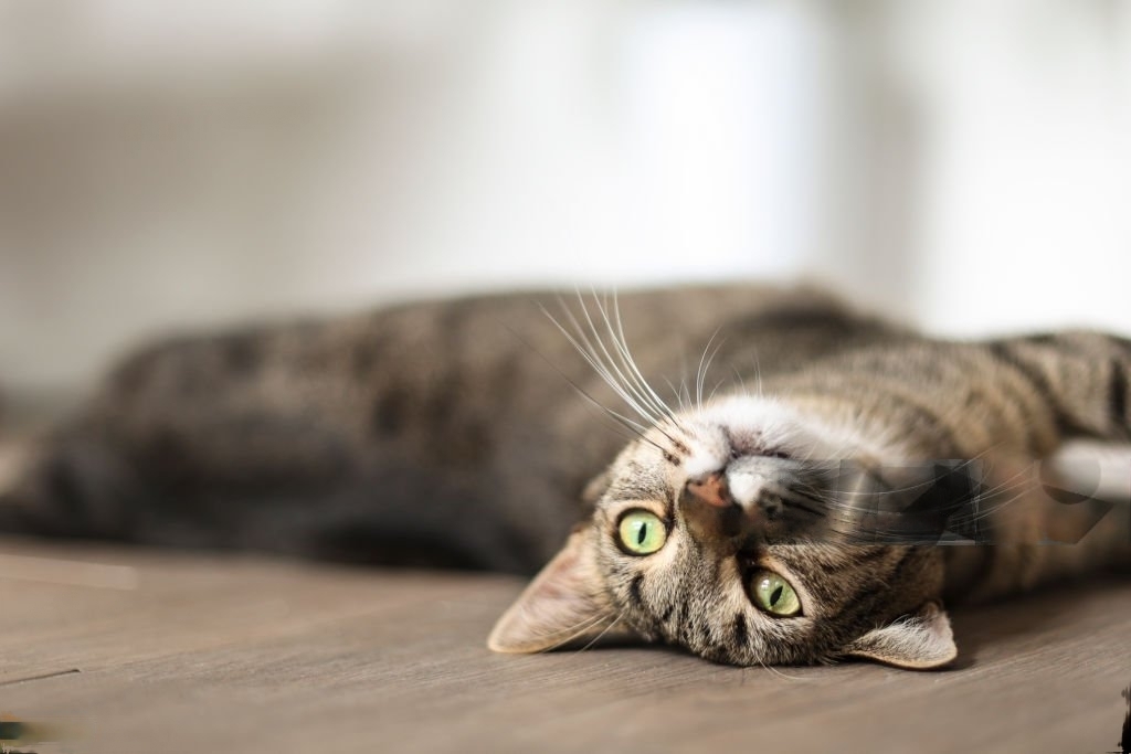
Feline Heat Cycle and the Importance of Spaying Female Cats When my cat enters heat? The feline heat cycle, also known as estrus, is a crucial aspect of a female cat’s reproductive behavior. Understanding when female cats enter heat and the importance of spaying can contribute significantly to their health and well-being. In this guide, we’ll explore the stages of the feline heat cycle and discuss the optimal time for spaying female cats. When Does a Female Cat Enter Heat? Female cats typically reach sexual maturity between the ages of five and nine months, although it can vary depending on factors such as breed, health, and environmental conditions. Once they reach puberty, female cats can enter their first heat cycle, signaling their readiness to mate and reproduce. The feline heat cycle consists of several stages, each characterized by specific behaviors and physiological changes: Proestrus: During the proestrus stage, which lasts approximately one to two days, female cats exhibit subtle signs of being in heat. They may become more affectionate towards their owners, vocalize more frequently, and display restless behavior. However, they are not yet receptive to mating. Estrus: The estrus stage, also known as the “true” heat cycle, is when female cats are fertile and receptive to mating. This phase typically lasts around five to seven days but can vary depending on individual cats. During estrus, female cats may display more pronounced behavioral changes, including increased vocalization, rolling on the floor, raising their hindquarters, and rubbing against objects or people. Additionally, they may attract male cats with their scent markings and vocalizations. Metestrus and Diestrus: After the estrus stage, female cats enter metestrus and diestrus, during which they are no longer receptive to mating. These stages mark the end of the heat cycle, and if the cat has not mated, she will gradually return to her normal behavior until the next heat cycle begins. The Best Time for Spaying Female Cats Spaying, also known as ovariohysterectomy, is the surgical removal of a female cat’s ovaries and uterus. Spaying offers numerous health benefits and helps prevent unwanted pregnancies, overpopulation, and certain reproductive-related health issues in female cats. The ideal time for spaying female cats is before they reach sexual maturity and enter their first heat cycle. Spaying at a young age, typically between eight and sixteen weeks old, is recommended by veterinary professionals and animal welfare organizations. Early spaying not only prevents unwanted litters but also reduces the risk of certain health problems, including mammary tumors, uterine infections (pyometra), and ovarian and uterine cancers. By spaying female cats before their first heat cycle, owners can also avoid the behavioral changes and challenges associated with heat cycles, such as yowling, urine spraying, and increased attempts to escape outdoors in search of mates. Additionally, early spaying contributes to population control efforts by preventing the birth of unwanted kittens, many of which end up in animal shelters or as strays. While spaying can be performed at any age, including during or after a cat’s first heat cycle, early spaying is generally considered the most beneficial option in terms of both health and behavior. However, even if a female cat has already experienced multiple heat cycles, spaying can still offer significant health benefits and help prevent future reproductive-related issues. Any other doubt? We are here to help, Contact us now. Contact Us Conclusion understanding the feline heat cycle and the importance of spaying female cats are essential aspects of responsible cat ownership. Female cats typically enter their first heat cycle between five and nine months of age, with the estrus stage marking their peak fertility. Spaying before the first heat cycle, ideally between eight and sixteen weeks old, offers numerous health benefits and helps prevent unwanted pregnancies and reproductive-related health issues. By spaying female cats at a young age, owners can promote their pets’ health and well-being while contributing to population control efforts and reducing the number of homeless cats in shelters and communities. Related Services Preventive Medicine Click here Pet Surgery Click here Alternative Medicine Click here Pet Dentistry Click here

Food allergies in dogs
Food allergies in dogs Recurrent otitis, occasional itchiness, and diarrhea? Food allergies in dogs are more common than many pet owners realize, affecting their furry companions’ well-being and quality of life. Just like humans, dogs can develop adverse reactions to certain ingredients in their food, leading to a range of symptoms from mild discomfort to severe health issues. Identifying and managing these allergies is crucial for ensuring the health and happiness of our canine friends. One essential tool in this process is the use of prescription diets for diagnostic purposes. Signs of Food Allergies: Recognizing the signs of food allergies in dogs is the first step in addressing this health concern. While symptoms can vary from one dog to another, some common signs of food allergies include: Skin Problems: Itching, scratching, redness, inflammation, and recurrent ear infections are often signs of food allergies in dogs. Persistent licking or chewing of the paws and skin lesions may also indicate an allergic reaction. Gastrointestinal Issues: Vomiting, diarrhea, flatulence, and frequent bowel movements can be symptoms of food allergies. Some dogs may also experience abdominal discomfort, bloating, or changes in appetite. Respiratory Symptoms: In severe cases, food allergies can manifest as respiratory issues such as coughing, wheezing, or difficulty breathing. These symptoms may indicate a more serious allergic reaction and require immediate veterinary attention. Ear Infections: Chronic or recurrent ear infections, characterized by redness, discharge, odor, and discomfort, are often linked to food allergies in dogs. Addressing the underlying allergy can help prevent future ear problems. Behavioral Changes: Dogs suffering from food allergies may exhibit changes in behavior, including irritability, restlessness, lethargy, or aggression. These behavioral changes can be a result of discomfort or pain caused by allergic reactions. Allergy Testing: Diagnosing food allergies in dogs typically involves a combination of clinical evaluation, dietary trials, and allergy testing. While elimination diet trials are considered the gold standard for diagnosing food allergies, allergy testing can provide valuable insights into potential allergens and guide the diagnostic process. Skin Testing: Skin prick tests or intradermal tests can be performed by veterinary dermatologists to identify potential allergens. Small amounts of common food allergens are injected into the dog’s skin, and any resulting reactions are observed. While skin testing can be informative, it may not always accurately reflect the dog’s true sensitivities. Blood Tests: Serum allergy testing, also known as serologic testing or IgE testing, measures the dog’s immune response to specific allergens by analyzing blood samples. While blood tests can provide useful information about potential allergens, they are not always definitive and may produce false positives or false negatives. Elimination Diet Trials: The most reliable method for diagnosing food allergies in dogs is through elimination diet trials. These trials involve feeding the dog a hypoallergenic diet consisting of novel or hydrolyzed proteins for a period of 8 to 12 weeks while monitoring for improvement in symptoms. If the dog’s symptoms resolve on the elimination diet and return upon reintroducing specific ingredients, it confirms a food allergy diagnosis. Any other doubt? We are here to help, Contact us now. Contact Us Conclusion In conclusion, recognizing the signs of food allergies and conducting appropriate allergy testing are essential steps in diagnosing and managing this common health concern in dogs. Prescription diets play a crucial role in the diagnostic process by providing novel or hydrolyzed proteins and eliminating potential allergens from the dog’s diet. By working closely with veterinarians and following their guidance, pet owners can identify the underlying causes of food allergies and develop tailored treatment plans to ensure the health and well-being of their canine companions. Related Services Preventive Medicine Click here Pet Surgery Click here Alternative Medicine Click here Pet Dentistry Click here

Periodontal desease
Periodontal Disease in Cats and Dogs Why my cat or dog has bad breath? Periodontal disease is a common dental condition that affects cats and dogs of all ages. It is characterized by inflammation and infection of the tissues surrounding the teeth, including the gums, periodontal ligament, and alveolar bone. Left untreated, periodontal disease can lead to pain, tooth loss, and systemic health issues. Understanding the causes, symptoms, and treatment options for periodontal disease is crucial for maintaining the oral health and overall well-being of our furry companions. Causes of Periodontal Disease Periodontal disease in cats and dogs typically begins with the accumulation of plaque, a sticky film of bacteria, saliva, and food particles that forms on the teeth. If not removed through regular brushing or professional dental cleanings, plaque can harden into tartar (dental calculus), which provides a breeding ground for bacteria. Over time, the bacteria in plaque and tartar can cause inflammation of the gums (gingivitis) and damage to the surrounding tissues, leading to periodontal disease. Several factors can contribute to the development of periodontal disease in cats and dogs, including: Poor Dental Hygiene: Lack of regular brushing and dental care can allow plaque and tartar to accumulate on the teeth, increasing the risk of periodontal disease. Diet: A diet high in carbohydrates or sticky foods can contribute to plaque formation and dental decay. Genetics: Some animals may be predisposed to developing periodontal disease due to genetic factors. Age: Older pets are more prone to periodontal disease, as years of plaque accumulation can take their toll on dental health. Symptoms of Periodontal Disease The signs of periodontal disease in cats and dogs can be subtle and may go unnoticed by pet owners until the disease has progressed. Common symptoms include: Bad Breath (Halitosis): Foul-smelling breath is often one of the first signs of periodontal disease and is caused by the presence of bacteria in the mouth. Gingivitis: Inflammation and redness of the gums may be visible, especially along the gumline. Dental Calculus: Yellow or brown tartar deposits may accumulate on the teeth, particularly near the gumline. Gum Recession: As periodontal disease progresses, the gums may recede, exposing the roots of the teeth. Loose or Missing Teeth: Advanced periodontal disease can cause tooth mobility or loss due to damage to the supporting structures. Diagnosis and Treatment Diagnosing periodontal disease in cats and dogs typically requires a thorough dental examination performed by a veterinarian. This may involve sedation or anesthesia to allow for a comprehensive assessment of the teeth and gums. Treatment for periodontal disease may include: Professional Dental Cleaning: Scaling and polishing the teeth to remove plaque and tartar buildup is a crucial step in managing periodontal disease. Tooth Extraction: Severely affected teeth may need to be extracted to prevent further damage to surrounding tissues. Antibiotics: In cases of advanced periodontal disease with bacterial infection, antibiotics may be prescribed to help control the infection. Home Dental Care: Regular tooth brushing, dental chews, and specialized dental diets can help prevent plaque buildup and maintain oral health between professional cleanings. Prevention Preventing periodontal disease in cats and dogs involves proactive dental care and regular veterinary check-ups. Pet owners can take the following steps to help maintain their pet’s oral health: Brushing: Daily tooth brushing with a pet-safe toothpaste and toothbrush can help remove plaque and prevent tartar buildup. Dental Treats and Toys: Providing dental treats and toys designed to promote chewing and reduce plaque can help support oral hygiene. Regular Veterinary Examinations: Annual or biannual dental check-ups with a veterinarian are essential for early detection and treatment of periodontal disease. Professional Dental Cleanings: Regular professional dental cleanings under anesthesia are recommended to remove plaque and tartar from hard-to-reach areas. Any other doubt? We are here to help, Contact us now. Contact Us Conclusion Periodontal disease is a common and preventable condition that can have serious consequences for the oral health and overall well-being of cats and dogs. By understanding the causes, symptoms, and treatment options for periodontal disease, pet owners can take proactive steps to maintain their pet’s dental health and quality of life. Regular dental care, both at home and with the guidance of a veterinarian, is key to preventing and managing periodontal disease in our furry companions. Related Services Preventive Medicine Click here Pet Surgery Click here Alternative Medicine Click here Pet Dentistry Click here

Lipomas in Dogs and Cats: Causes, Symptoms, and Treatment
Lipomas in Dogs and Cats: Causes, Symptoms, and Treatment What is a lipoma? Lipomas, commonly known as fatty tumors, are benign growths that develop underneath the skin in dogs and cats. While they are usually harmless, their presence can cause concern for pet owners. Understanding lipomas, including their causes, symptoms, and treatment options, is essential for ensuring the health and well-being of our furry companions. What are Lipomas? Lipomas are soft, movable masses that typically develop in the subcutaneous tissue, which is the layer of tissue just beneath the skin. These growths consist of fat cells and often feel like rubbery lumps under the skin. Lipomas can vary in size from small pea-sized nodules to larger masses that can grow several inches in diameter. Causes of Lipomas The exact cause of lipomas in dogs and cats is not fully understood. However, several factors may contribute to their development: Genetics: Certain breeds, such as Labrador Retrievers, Doberman Pinschers, and Dachshunds, are more prone to developing lipomas. Age: Lipomas are more common in older pets, typically appearing in dogs and cats around middle age or older. Obesity: Overweight or obese pets may have a higher risk of developing lipomas, as excess fat tissue can contribute to their formation. Hormonal Imbalance: Hormonal changes or imbalances may play a role in the development of lipomas, although more research is needed in this area. Any other doubt? We are here to help, Contact us now. Contact Us Symptoms of Lipomas In many cases, lipomas do not cause any symptoms and are often discovered during routine physical examinations by a veterinarian. However, some pets may exhibit the following signs: Visible Lumps: Owners may notice soft, round lumps beneath the skin, which can vary in size and number. Limping or Discomfort: If a lipoma grows large enough to press on nearby nerves or tissues, it may cause limping or discomfort in affected pets. Changes in Mobility: Pets with lipomas located in areas that interfere with movement, such as the limbs or torso, may exhibit changes in mobility or range of motion. Diagnosis and Treatment To diagnose a lipoma, a veterinarian will perform a physical examination and may recommend additional tests, such as fine needle aspiration or biopsy, to confirm the diagnosis and rule out other potential causes. In most cases, treatment for lipomas in dogs and cats is not necessary unless they are causing discomfort or interfering with the pet’s quality of life. However, if a lipoma is large, rapidly growing, or causing other health problems, surgical removal may be recommended. During surgery, the veterinarian will remove the lipoma along with a margin of healthy tissue to prevent recurrence. In some cases, liposuction may be used as a less invasive method of removal. Preventative Measures While it may not be possible to prevent lipomas entirely, there are steps pet owners can take to help reduce the risk of their development: Maintain a Healthy Weight: Keeping pets at a healthy weight through proper diet and regular exercise can help prevent the accumulation of excess fat tissue. Regular Veterinary Examinations: Routine veterinary check-ups can help detect lipomas early and monitor their growth over time. Monitor for Changes: Pet owners should regularly check their pets for any new lumps or bumps and report any changes to their veterinarian promptly. Conclusion Lipomas are common benign growths that can develop in dogs and cats, particularly as they age. While they are usually harmless, it’s essential for pet owners to be aware of the signs and symptoms associated with lipomas and to seek veterinary care if any concerns arise. With proper diagnosis and management, pets with lipomas can lead happy and healthy lives. Preventive Medicine Click here Pet Surgery Click here Alternative Medicine Click here Pet Dentistry Click here

Feline triaditis
Understanding Feline Triaditis: Causes, Diagnosis, and Treatment What is happening to my cat? Feline triaditis is a complex syndrome characterized by the concurrent inflammation of three major organs in cats: the liver, pancreas, and small intestine. This condition presents significant challenges in diagnosis and management due to its multifactorial nature and overlapping clinical signs. Understanding the causes, diagnostic approach, and treatment options for feline triaditis is crucial for veterinarians and cat owners alike. Causes The exact cause of feline triaditis remains elusive, but it is believed to result from a combination of factors including: Inflammatory Response: The inflammatory cascade triggered by various insults such as infections, dietary indiscretion, or immune-mediated processes can lead to concurrent inflammation of the liver, pancreas, and intestines. Bacterial Infections: Bacterial infections, particularly in the gastrointestinal tract, can contribute to the development of feline triaditis. Bacterial translocation from the intestines to the liver and pancreas may exacerbate inflammation. Immune-Mediated Factors: Immunological abnormalities and dysregulation of the immune response may play a role in the pathogenesis of feline triaditis, leading to chronic inflammation and tissue damage. Dietary Factors: Certain dietary components, including high-fat diets or food allergens, have been implicated in the development of pancreatitis and intestinal inflammation, contributing to the triaditis syndrome. Genetic Predisposition: Some cats may have a genetic predisposition to developing triaditis, although specific genetic markers have yet to be identified. Diagnostic Approach to Feline Triaditis: Diagnosing feline triaditis can be challenging due to its nonspecific clinical signs and overlapping manifestations with other diseases. A comprehensive diagnostic approach may include: Physical Examination: A thorough physical examination by a veterinarian may reveal signs of abdominal pain, jaundice, dehydration, or other systemic abnormalities suggestive of triaditis. Blood Tests: Blood tests, including a complete blood count (CBC), serum biochemistry profile, and specific markers such as pancreatic lipase immunoreactivity (PLI) and liver enzymes (ALT, ALP), can help assess organ function and identify abnormalities indicative of triaditis. Imaging Studies: Abdominal ultrasound is a valuable diagnostic tool for evaluating the liver, pancreas, and intestines, detecting abnormalities such as inflammation, thickening of the intestinal wall, or the presence of gallstones. Fecal Analysis: Fecal examination may be performed to rule out parasitic infections or assess for the presence of inflammatory markers such as fecal elastase. Diagnostic Imaging: Additional imaging modalities such as contrast-enhanced computed tomography (CT) or magnetic resonance imaging (MRI) may be utilized in complex cases to evaluate the extent of organ involvement further and guide treatment decisions. Any other doubt? We are here to help, Contact us now. Contact Us Treatment Options The treatment of feline triaditis aims to alleviate clinical signs, manage complications, and address underlying causes. Treatment modalities may include: Supportive Care: Supportive care is essential for cats with triaditis and may include intravenous fluid therapy to correct dehydration, nutritional support, pain management, and monitoring for complications such as hepatic encephalopathy or pancreatitis. Dietary Management: A highly digestible, low-fat diet may be recommended to reduce pancreatic stimulation and minimize gastrointestinal inflammation. Prescription diets formulated for cats with pancreatitis or gastrointestinal sensitivity may be beneficial. Medications: Depending on the underlying cause and severity of inflammation, medications such as antibiotics, anti-inflammatory drugs (e.g., corticosteroids), or immunosuppressive agents may be prescribed to control inflammation and modulate the immune response. Treatment of Complications: Complications such as hepatic lipidosis, sepsis, or secondary infections may require specific interventions, including antibiotics, nutritional support, or intensive care. Monitoring and Follow-up: Regular monitoring of clinical signs, laboratory parameters, and imaging studies is essential to assess treatment response, detect recurrence, and adjust the treatment plan as needed. Follow-up appointments with a veterinarian are crucial for long-term management and supportive care. In conclusion, feline triaditis is a complex syndrome characterized by inflammation of the liver, pancreas, and small intestine in cats. While the exact cause remains unclear, a multifactorial interplay of inflammatory, infectious, dietary, and immune-mediated factors is believed to contribute to its development. Diagnosing feline triaditis requires a thorough diagnostic evaluation encompassing physical examination, blood tests, imaging studies, and fecal analysis. Treatment options focus on supportive care, dietary management, medications, and addressing underlying causes to improve clinical outcomes and enhance the quality of life for affected cats. Early recognition, prompt intervention, and ongoing monitoring are essential for managing feline triaditis effectively and minimizing complications. Related Services Preventive Medicine Click here Pet Surgery Click here Alternative Medicine Click here Pet Dentistry Click here
