
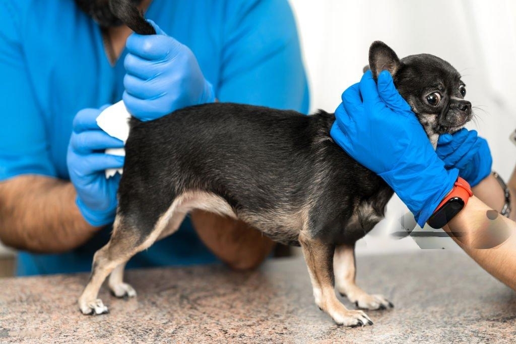
Anal Glands in Dogs: Pathologies and Treatments What are the anal glands? Anal glands, also known as anal sacs, are small, paired structures located on either side of a dog’s anus. These glands are responsible for secreting a fluid that is used for marking territory and communication with other dogs. While these glands serve an important biological function, they can become a source of discomfort and medical issues if they are not functioning properly. Understanding the anatomy, common pathologies, and treatment options for anal gland problems is crucial for maintaining your dog’s health and well-being. Anatomy and Function Each anal gland is situated at approximately 4 o’clock and 8 o’clock positions around the anus. These glands secrete a foul-smelling fluid that is expelled during defecation or, in some cases, through voluntary contraction. The secretion is unique to each dog, which helps in identifying individual animals and marking territory. Common problems: Anal Gland Impaction Description: Anal gland impaction occurs when the fluid within the glands becomes thickened or the ducts become blocked. This prevents the glands from emptying naturally. Symptoms: Dogs with impaction often show signs of discomfort, such as scooting their rear end on the ground, excessive licking of the anal area, or signs of pain when the area is touched. The fluid can also become more viscous and may lead to discomfort or a noticeable bulge around the anus. Treatment: Mild cases of impaction may be managed through regular expression of the glands by a veterinarian. In more severe cases, treatment might involve the use of warm compresses and manual expression of the glands. If the problem persists, the veterinarian may recommend dietary changes or medications to help regulate gland function and fluid consistency. Anal Gland Infection Description: Bacterial infections can occur when the impaction of anal glands is not addressed. The infection can cause swelling, pain, and discharge that may be foul-smelling. Symptoms: Dogs with anal gland infections typically exhibit signs of pain, such as vocalizing or biting at their rear end. There may be redness, swelling, and an unpleasant odor coming from the anal area. In severe cases, fever and lethargy can also be observed. Treatment: Treatment involves antibiotics to clear the infection and pain relief medications to manage discomfort. The veterinarian may also need to perform a thorough cleaning of the infected glands and provide follow-up care to ensure the infection resolves completely. Anal Gland Abscess Description: An abscess forms when an infection within the anal gland progresses and leads to the formation of a pus-filled pocket. This is a more severe complication of untreated infections or impactions. Symptoms: Dogs with anal gland abscesses will show pronounced pain, swelling, and redness. The area may be hot to the touch, and the dog may have difficulty sitting or walking. Pus may be visible if the abscess ruptures. Treatment: Surgical intervention is often required to drain the abscess and address the infection. The dog may need to be placed on antibiotics and pain relief medications post-surgery. Maintaining good hygiene and regular follow-up care is essential to prevent recurrence. Anal Gland Tumors Description: Although less common, tumors can develop in the anal glands, with potential types including both benign and malignant forms. These tumors may cause a range of symptoms depending on their size and malignancy. Symptoms: Symptoms can be similar to those of impaction or infection but may also include unusual lumps near the anus, persistent bleeding, or changes in bowel habits. Weight loss and lethargy might occur in advanced cases. Treatment: Diagnosis typically involves imaging studies and biopsy to determine the nature of the tumor. Treatment options include surgical removal of the tumor, radiation therapy, and sometimes chemotherapy. The prognosis depends on the type and stage of the tumor, and a tailored treatment plan is essential for effective management. Preventive Measures and Long-Term Management To prevent recurring anal gland problems, consider the following strategies: Diet and Nutrition: A high-fiber diet can help improve the consistency of stool, which can naturally help with the expression of anal glands. Consult your veterinarian for dietary recommendations that suit your dog’s specific needs. Regular Veterinary Check-ups: Routine vet visits can help monitor the health of your dog’s anal glands and address any early signs of issues. Regular anal gland expressions by a veterinarian may be recommended for dogs prone to problems. Maintaining Hygiene: Keeping the anal area clean and dry can help prevent infections and irritations. Regular grooming and bathing can also contribute to overall anal gland health. Conclusion Anal gland problems in dogs, while often manageable, require prompt attention to prevent complications. Understanding the anatomy and potential issues related to anal glands allows pet owners to recognize symptoms early and seek appropriate veterinary care. With proper treatment and preventive measures, many anal gland issues can be resolved effectively, ensuring your furry friend remains comfortable and healthy. Regular veterinary visits, a balanced diet, and good hygiene practices play a crucial role in maintaining anal gland health and overall well-being for your canine companion. Any other doubt? We are here to help, Contact us now. Contact Us
Osteosarcoma the bone cancer
Osteosarcoma: bone cancer What osteosarcoma? Osteosarcoma is a malignant bone tumor that primarily affects dogs and, less commonly, cats. This aggressive cancer originates in the bone’s cells and can lead to severe health complications if not promptly addressed. Understanding the breeds most susceptible to osteosarcoma, the available treatments, and the importance of monitoring for metastasis is crucial for effective management of this condition. Breeds at RiskDogs: Osteosarcoma is most prevalent in large and giant dog breeds. Breeds such as Great Danes, Rottweilers, Doberman Pinschers, and Saint Bernards are particularly prone to developing this aggressive bone cancer. These breeds often face a higher risk due to their rapid growth rates and large skeletal structure. Additionally, breeds like Labrador Retrievers and German Shepherds also show a predisposition, although to a lesser extent compared to the aforementioned giants. Cats: Osteosarcoma is considerably rarer in cats, but certain breeds may still exhibit a higher susceptibility. Maine Coons, with their large size and robust build, and Siamese cats, known for their unique genetic predispositions, are among those that can be affected. However, the overall incidence in cats is much lower compared to dogs. Symptoms and DiagnosisThe clinical presentation of osteosarcoma in pets often begins with localized pain and swelling, which might be mistaken for less serious conditions. Common symptoms include lameness, reluctance to use a limb, and observable swelling around the affected bone. In advanced cases, pets may also exhibit signs of systemic illness such as lethargy and weight loss. Diagnosis typically involves a combination of physical examination, radiographic imaging, and sometimes biopsy. X-rays are crucial for visualizing the tumor’s presence and extent. An X-ray will often show an aggressive, irregular bone lesion that might be accompanied by a noticeable periosteal reaction (new bone formation around the tumor). Treatment OptionsSurgery: The primary treatment for osteosarcoma in dogs and cats is surgical amputation of the affected limb. Although this might sound drastic, it often provides the best chance for long-term survival, especially when performed before the cancer has spread. Amputation is generally followed by a rehabilitation period where pets adjust to life on three legs, which they typically adapt to surprisingly well. Chemotherapy: Post-surgical chemotherapy is commonly recommended to address potential metastasis, especially to the lungs. Chemotherapy drugs such as doxorubicin and carboplatin are frequently used to target any remaining cancer cells that might have dispersed from the primary tumor site. While chemotherapy can have side effects like gastrointestinal upset or temporary suppression of bone marrow function, it plays a critical role in improving survival rates. Radiation Therapy: In cases where amputation is not possible or as an adjunct to surgery, radiation therapy can be employed to target and shrink the tumor. This treatment is generally used for palliative care to alleviate pain and improve the quality of life for pets with osteosarcoma, particularly when the tumor is not amenable to surgical removal. Monitoring for MetastasisOne of the major concerns with osteosarcoma is its propensity to metastasize, most commonly to the lungs. Metastasis occurs when cancer cells break away from the primary bone tumor and travel through the bloodstream to form secondary tumors in other organs. To detect and address metastasis early, regular chest X-rays are essential. Chest X-rays help in monitoring the lungs for any signs of metastatic spread. Since osteosarcoma cells can quickly invade the lungs, these imaging studies are critical for determining the disease’s progression and guiding treatment decisions. Early detection of metastasis allows for timely intervention, whether through additional chemotherapy, supportive care, or other strategies to manage the cancer’s spread. ConclusionOsteosarcoma is a severe and aggressive cancer affecting both dogs and cats, with large and giant dog breeds being particularly vulnerable. The primary treatment strategy includes surgical amputation of the affected limb, followed by chemotherapy to address possible metastasis. Radiation therapy can be used as a supplementary treatment or for palliative care. Due to the high likelihood of metastasis, regular chest X-rays are vital for monitoring and managing the disease effectively. Pet owners should be vigilant for symptoms and work closely with their veterinarians to ensure comprehensive care and optimal outcomes for their beloved companions. What is happening? Reverse sneezing involves sudden, repeated inhalations through the nose, resulting in a snorting or honking sound. Triggers can include excitement, irritants, allergies, infections, pulling on a leash, or anatomical factors. Small breeds like York shire terriers, chihuahuenios, poodles, and bulldogs are more prone to develop this problem but that doesn’t exclude mixed breeds or bigger breeds like golden retrievers and grate and Newfoundland for having this coughing-like event. Symptoms: Rapid, noisy inhalations through the nose, accompanied by a stiffening of the body, and the dog extending its head and neck. Duration: Episodes typically last for a few seconds to a minute, resolving on their own. Reverse sneezing differs from regular sneezing or coughing and usually doesn’t require medical intervention. What can I do? If you find your furry friend caught in a reverse sneezing fit, there are a few techniques you can try to help alleviate the episode. Massaging the throat gently, covering the nostrils momentarily to encourage swallowing, or offering a small amount of water can sometimes help shorten these episodes and offer relief to your pup. However, it’s important to note that while occasional reverse sneezing is generally harmless, frequent or severe episodes warrant a consultation with a veterinarian. If these episodes become more frequent or are accompanied by other concerning symptoms, seeking professional advice becomes crucial. Veterinarians can provide insight into understanding and effectively managing this condition, ensuring your dog’s well-being. While reverse sneezing is common and often not a cause for alarm, it’s essential to remember that any coughing-like event or unusual respiratory behavior in dogs should prompt veterinary attention. A professional diagnosis ensures accurate identification and appropriate management of the underlying cause. Fun Fact Incredible Scent Receptors: Dogs possess an extraordinary olfactory system with around 300 million scent receptors. This number is significantly higher than the mere 5-6 million scent receptors found in humans. This incredible number
Luxating patella
Luxating patella What is Luxating patella? Luxating patella, commonly known as kneecap dislocation, is a prevalent orthopedic condition in dogs, particularly small and toy breeds. It occurs when the patella, or kneecap, slips out of its normal position within the groove of the femur (thigh bone), leading to discomfort, lameness, and progressive joint damage if left untreated. Understanding the stages and surgical treatments associated with luxating patella is crucial for managing this condition effectively. Stages of Luxating Patella: Luxating patella is categorized into four stages based on the severity and frequency of displacement: Grade 1 (Mild): In Grade 1, the patella can be manually luxated (moved out of place) but returns to its normal position when released. Dogs with Grade 1 luxation may show occasional lameness or favoring of the affected limb, particularly after physical activity. At this stage, the condition may not be causing significant pain or joint degeneration. Grade 2 (Moderate): Grade 2 involves more frequent patellar luxation, where the kneecap slips out of place with moderate ease and may spontaneously return to the groove of the femur. Lameness becomes more noticeable and can occur more frequently, especially during exercise or when the dog jumps or runs. Over time, Grade 2 luxation can lead to progressive joint changes such as cartilage wear and potential early signs of arthritis. Grade 3 (Severe): In Grade 3 luxation, the patella is frequently out of position and often remains luxated until manually corrected. Dogs with Grade 3 luxation typically exhibit persistent lameness and discomfort. They may hold the affected limb up or avoid using it altogether. Joint changes are more pronounced at this stage, with increased risk of cartilage damage, inflammation, and worsening arthritis. Grade 4 (Permanent): Grade 4 is the most severe stage, where the patella remains permanently luxated and cannot be manually repositioned. Dogs with Grade 4 luxation experience constant pain, significant lameness, and severe joint degeneration. This stage often requires prompt surgical intervention to alleviate pain and improve joint function. Surgical Treatment Options: Surgical correction is often recommended for Grades 2 to 4 luxating patella cases, depending on the severity and the dog’s overall health. The goal of surgery is to realign the patella and deepen the femoral groove to prevent further displacement. Common surgical techniques include: Trochlear Block (Trochleoplasty): This procedure involves deepening the groove in which the patella sits (trochlea) on the femur. By creating a more stable groove, the patella is less likely to slip out of place, reducing the risk of recurrence. Trochlear block can be performed alone or in combination with other surgical techniques depending on the specific needs of the dog. Tibial Tuberosity Transposition (TTT): TTT involves repositioning the attachment of the patellar ligament on the tibia, effectively realigning the patella to prevent luxation. This procedure aims to correct the anatomical abnormalities that contribute to patellar instability. TTT is often recommended for more severe cases where trochlear block alone may not be sufficient. Lateral Imbrication: Lateral imbrication involves tightening the joint capsule and soft tissues on the lateral side of the knee joint. This technique helps to support the patella and prevent lateral luxation. Lateral imbrication is typically performed in combination with other procedures for comprehensive stabilization. Medial Patellar Luxation Correction: For dogs with medial patellar luxation (where the patella moves towards the inside of the knee), corrective surgery focuses on realigning the patella to its correct position. Surgical techniques may involve releasing tight tissues on the inside of the knee and adjusting the position of the attachment of the patellar ligament. Post-Surgical Care: Post-operative care is essential for the successful recovery of dogs undergoing luxating patella surgery. It typically includes: Restricted Activity: Limiting physical activity and providing a controlled environment to prevent excessive stress on the surgical site. Physical Therapy: Gentle exercises to improve range of motion and strength gradually. Pain Management: Medications to manage post-operative pain and inflammation. Follow-up Examinations: Regular veterinary check-ups to monitor healing progress and address any complications. Conclusion: Luxating patella is a common orthopedic issue in dogs that can significantly impact their quality of life if not properly managed. Understanding the stages of luxation and the surgical treatment options available is crucial for pet owners and veterinarians to make informed decisions regarding the best course of action for affected dogs. Early detection and intervention can help prevent irreversible joint damage and alleviate pain, allowing dogs to lead active and comfortable lives post-surgery Any other doubt? We are here to help, Contact us now. Contact Us
Kidney disease in dogs
Understanding Kidney disease and SDMA: A Game-Changing Biomarker Kidney disease? Recent advancements in veterinary medicine have highlighted the importance of Symmetric Dimethylarginine (SDMA) as a biomarker for kidney function. SDMA is a molecule that is excreted by the kidneys, and its blood levels correlate closely with the glomerular filtration rate (GFR), which is a measure of kidney function. Unlike traditional markers such as creatinine, SDMA can detect kidney disease earlier, often when only 25-40% of kidney function has been lost. This early detection is crucial because it allows veterinarians to intervene sooner, potentially slowing the progression of the disease and improving the prognosis for affected dogs. Stages of Kidney Disease: IRIS Classification The International Renal Interest Society (IRIS) has developed a staging system to classify the severity of chronic kidney disease in dogs based on blood creatinine levels, blood pressure, urine protein levels, and other parameters. This staging system helps veterinarians to better understand the progression of the disease and tailor treatment plans accordingly: Stage 1 (Mild): Dogs in this stage have mild kidney disease with creatinine levels slightly above normal. Often, there are no clinical signs of kidney disease apparent at this stage. Stage 2 (Mild to Moderate): Kidney function is moderately decreased, and dogs may start showing mild symptoms such as increased thirst and urination. Stage 3 (Moderate): Dogs in stage 3 have moderately severe kidney disease, with significant elevation in creatinine levels. Symptoms may include weight loss, poor appetite, and lethargy. Stage 4 (Severe): This is the most advanced stage of kidney disease, characterized by a marked decrease in kidney function. Dogs may exhibit severe clinical signs such as vomiting, diarrhea, and severe dehydration. Clinical Signs and Diagnosis Recognizing the signs of kidney disease early is crucial for prompt diagnosis and treatment. Common clinical signs include increased thirst and urination, decreased appetite, weight loss, vomiting, and lethargy. However, these signs can be nonspecific and easily attributed to other health conditions, highlighting the importance of routine veterinary check-ups and blood work. Diagnosing kidney disease typically involves a combination of blood tests (including SDMA and creatinine levels), urine analysis, and sometimes imaging techniques such as ultrasound to evaluate the size and structure of the kidneys. Treatment and Management While chronic kidney disease cannot be cured, early intervention and appropriate management can significantly improve a dog’s quality of life and slow disease progression. Treatment may include dietary management (such as prescription kidney diets that are lower in protein and phosphorus), medications to control blood pressure and manage symptoms, fluid therapy to maintain hydration, and supportive care as needed. Regular monitoring of kidney function through blood tests and urine analysis is essential to adjust treatment plans as the disease progresses. Dogs with more advanced stages of kidney disease may require more intensive management and supportive care to maintain their health and well-being. Importance of Nutritional Support Nutrition plays a critical role in managing kidney disease in dogs. Prescription kidney diets are formulated to support kidney function by reducing the workload on the kidneys and minimizing the buildup of waste products in the bloodstream. These diets are typically lower in protein and phosphorus, which helps to reduce the strain on the kidneys and slow the progression of the disease. Supplements such as omega-3 fatty acids and antioxidants may also be beneficial in supporting kidney health and reducing inflammation. It is essential for dog owners to work closely with their veterinarian to develop a tailored nutrition plan that meets the specific needs of their dog based on their stage of kidney disease and overall health status. Prognosis and Long-Term Care The prognosis for dogs with kidney disease varies depending on the stage of the disease at diagnosis, the underlying cause, and how well the dog responds to treatment. With early detection and comprehensive management, many dogs can live for months to years with a good quality of life. However, it is essential to recognize that kidney disease is progressive, and ongoing monitoring and adjustments to the treatment plan are often necessary to maintain stability and manage complications as they arise. In conclusion, kidney disease is a common and serious health condition in dogs, particularly as they age. The advent of SDMA as a biomarker has revolutionized early detection and monitoring, allowing veterinarians to intervene sooner and improve outcomes for affected dogs. Understanding the IRIS staging system helps veterinarians to classify the severity of chronic kidney disease and tailor treatment plans accordingly. With timely diagnosis, appropriate management, and ongoing veterinary care, many dogs with kidney disease can continue to enjoy a good quality of life for an extended period. Any other doubt? We are here to help, Contact us now. Contact Us
The heart: dogs and cats
The Heart : Cats and Dogs edition how this works? Understanding how the heart works in cats and dogs is essential for pet owners to ensure their pets remain healthy and happy. The heart is a vital organ that pumps blood throughout the body, delivering oxygen and nutrients to tissues and removing waste products. Anatomy of the Heart The heart of cats and dogs has four chambers: two atria (upper chambers) and two ventricles (lower chambers). These chambers work together to circulate blood efficiently. Here’s a breakdown of the heart’s components: Right Atrium: Receives deoxygenated blood from the body. Right Ventricle: Pumps deoxygenated blood to the lungs via the pulmonary artery. Left Atrium: Receives oxygenated blood from the lungs. Left Ventricle: Pumps oxygenated blood to the body through the aorta. In addition to the chambers, the heart contains valves that ensure blood flows in the correct direction. These valves are: Tricuspid Valve: Between the right atrium and right ventricle. Pulmonary Valve: Between the right ventricle and the pulmonary artery. Mitral Valve: Between the left atrium and left ventricle. Aortic Valve: Between the left ventricle and the aorta. Blood Circulation Process The process of blood circulation in cats and dogs involves two main circuits: the pulmonary circulation and the systemic circulation. Pulmonary Circulation: This circuit moves blood between the heart and the lungs. Deoxygenated blood from the body enters the right atrium. The right atrium contracts, pushing blood through the tricuspid valve into the right ventricle. The right ventricle contracts, sending blood through the pulmonary valve into the pulmonary artery, which carries it to the lungs. In the lungs, blood releases carbon dioxide and picks up oxygen. Systemic Circulation: This circuit moves blood between the heart and the rest of the body. Oxygenated blood from the lungs enters the left atrium. The left atrium contracts, pushing blood through the mitral valve into the left ventricle. The left ventricle contracts, sending blood through the aortic valve into the aorta, which distributes it throughout the body. As blood travels through the body, it delivers oxygen and nutrients to tissues and picks up waste products and carbon dioxide, eventually returning to the right atrium. Heartbeat Regulation The heartbeat in cats and dogs is controlled by an electrical conduction system. The main components of this system are: Sinoatrial (SA) Node: Located in the right atrium, the SA node is the natural pacemaker of the heart. It generates electrical impulses that cause the atria to contract. Atrioventricular (AV) Node: Located between the atria and ventricles, the AV node receives impulses from the SA node and passes them to the ventricles. Bundle of His and Purkinje Fibers: These fibers carry the impulses through the ventricles, causing them to contract and pump blood. Differences Between Cats and Dogs While the basic function of the heart is similar in both cats and dogs, there are some differences: Heart Rate: Dogs generally have a slower heart rate than cats. A typical heart rate for a dog is 60-140 beats per minute, depending on size and activity level. Cats typically have a heart rate of 140-220 beats per minute. Heart Size: In proportion to their body size, cats have smaller hearts compared to dogs. This means their hearts beat faster to maintain effective blood circulation. Common Heart Problems Cats and dogs can suffer from various heart conditions. Some common issues include: Heart Murmurs: These are abnormal sounds heard during a heartbeat, indicating turbulent blood flow. They can be harmless or indicate an underlying problem. Heart Disease: Conditions such as cardiomyopathy (disease of the heart muscle) and heart valve disease can affect both cats and dogs. Congestive Heart Failure: This condition occurs when the heart cannot pump blood effectively, leading to fluid buildup in the lungs and other tissues. Conclusion The heart is a vital organ in cats and dogs, ensuring that oxygen and nutrients are delivered throughout their bodies while waste products are removed. By understanding how the heart works, pet owners can better monitor their pets’ health and seek veterinary care if they notice signs of heart problems, such as coughing, difficulty breathing, or lethargy. Regular check-ups with a veterinarian can help detect and manage heart conditions early, promoting a longer, healthier life for pets. Any other doubt? We are here to help, Contact us now. Contact Us
Lungs Cats/Dogs
Understanding the Lungs : Cats and Dogs How theyb work? The respiratory systems of cats and dogs are quite similar and play a crucial role in keeping these animals healthy. Understanding how their lungs work can help pet owners recognize signs of respiratory problems early on and seek appropriate care. Anatomy of the Respiratory System Both cats and dogs have a respiratory system that includes the nose, mouth, trachea (windpipe), bronchi, bronchioles, and lungs. Here’s a simple breakdown of each component’s role: Nose and Mouth: These are the entry points for air. When a cat or dog breathes in, air is filtered and moistened by the nasal passages or mouth. Trachea: This is a tube that connects the throat to the lungs. It splits into two main bronchi, one for each lung. Bronchi and Bronchioles: The bronchi further divide into smaller tubes called bronchioles within the lungs. These small airways distribute air evenly throughout the lungs. Lungs: The lungs are the main organs of the respiratory system. They contain tiny air sacs called alveoli, where the exchange of oxygen and carbon dioxide takes place. Breathing Process The breathing process in cats and dogs involves two main actions: inhalation (breathing in) and exhalation (breathing out). Inhalation: When a cat or dog inhales, the diaphragm (a large muscle located below the lungs) contracts and moves downward. This creates a vacuum that pulls air into the lungs. The intercostal muscles (located between the ribs) also help by expanding the chest cavity. Exhalation: During exhalation, the diaphragm and intercostal muscles relax, pushing air out of the lungs. This process expels carbon dioxide, a waste product of metabolism, from the body. Gas Exchange The primary function of the lungs is to facilitate gas exchange. This occurs in the alveoli, which are surrounded by tiny blood vessels called capillaries. Here’s how it works: Oxygen Intake: When air reaches the alveoli, oxygen passes through the thin walls of the alveoli into the capillaries. Oxygen Transport: Hemoglobin in the red blood cells picks up the oxygen and carries it to the rest of the body. Carbon Dioxide Removal: Simultaneously, carbon dioxide, a waste product from the body’s metabolism, moves from the blood into the alveoli. During exhalation, this carbon dioxide is expelled from the body. Regulation of Breathing The breathing rate in cats and dogs is regulated by the brain, which constantly monitors the levels of carbon dioxide and oxygen in the blood. When the carbon dioxide level rises, the brain signals the respiratory muscles to increase the breathing rate. This ensures that enough oxygen is taken in and carbon dioxide is expelled efficiently. Differences Between Cats and Dogs While the basic function of the respiratory system is similar in both cats and dogs, there are some differences: Breathing Rate: Generally, dogs have a faster breathing rate compared to cats. The normal breathing rate for a dog is 10-35 breaths per minute, while for cats, it’s 20-30 breaths per minute. Panting: Dogs often pant to cool down, as they have fewer sweat glands than cats. Panting helps them release excess heat. Cats pant less frequently, usually only when stressed or overheated. Common Respiratory Problems Cats and dogs can suffer from various respiratory problems. Some common issues include: Infections: Viral or bacterial infections can lead to conditions like pneumonia or bronchitis. Asthma: More common in cats, asthma causes inflammation and narrowing of the airways. Allergies: Both cats and dogs can have allergic reactions that affect their breathing. Collapsed Trachea: More common in small dog breeds, this condition involves the weakening of the tracheal rings, causing difficulty breathing. Conclusion The lungs are vital for cats and dogs, ensuring they receive the oxygen needed for their bodies to function and removing waste gases. By understanding how their respiratory system works, pet owners can better care for their furry friends and recognize when something might be wrong. If a pet shows signs of respiratory distress, such as persistent coughing, wheezing, or difficulty breathing, it’s essential to seek veterinary care promptly. Any other doubt? We are here to help, Contact us now. Contact Us
Basic Liver Function in Cats and Dogs
Comprehensive Guide to Liver Function in Cats and Dogs Image from Freepik The liver is a vital organ that performs numerous essential functions in both cats and dogs. Understanding these functions is crucial for pet owners to ensure their furry friends’ overall health and well-being. This comprehensive guide delves into the liver’s key functions, its importance in cats and dogs, and tips for maintaining liver health. Key Functions of the Liver Detoxification: The liver helps remove toxins and waste products from the blood. This detoxification process is crucial for keeping the body free from harmful substances. By filtering out these toxins, the liver ensures that the blood circulating through the body is clean and safe, vital for overall health. Metabolism: The liver processes nutrients from pets’ food, converting these nutrients into energy and storing essential vitamins and minerals. This metabolic function supports the body’s energy needs and helps maintain a balanced nutritional state. The liver’s role in metabolism is fundamental to the health and vitality of both cats and dogs. Production of Bile: The liver produces bile, a substance that aids in the digestion of fats. Bile is stored in the gallbladder and released into the intestines during digestion. This function is essential for the efficient breakdown and absorption of dietary fats, contributing to proper digestive health. Protein Synthesis: The liver produces important proteins necessary for blood clotting and other vital functions. These proteins include albumin, which helps maintain blood volume and pressure, and clotting factors essential for preventing excessive bleeding. Protein synthesis by the liver is crucial for maintaining homeostasis and supporting various bodily functions. Immune System Support: The liver helps fight infections by removing bacteria and other pathogens from the blood. This immune function protects the body against infections and maintains a robust immune response. The liver’s role in immune support is critical to the body’s defense mechanism. Liver Health in Cats and Dogs Maintaining liver health is essential for the overall well-being of pets. Both cats and dogs can suffer from liver diseases, but the causes and manifestations can differ between species. Cats: Cats are particularly prone to liver diseases such as hepatic lipidosis (fatty liver disease), which often occurs if they stop eating suddenly. This condition can be severe and requires prompt veterinary intervention. Proper nutrition and regular veterinary check-ups are crucial to maintaining liver health in cats. Ensuring that cats receive a balanced diet and adequate hydration can help prevent liver issues. Monitoring their eating habits and seeking veterinary care if they stop eating is essential for early detection and treatment of liver problems. Dogs: Dogs can suffer from various liver diseases, including chronic hepatitis and liver tumors. These conditions can significantly impact a dog’s health and quality of life. Regular exercise, a balanced diet, and routine health screenings can help keep a dog’s liver healthy. Providing dogs with a diet rich in essential nutrients and ensuring they get regular physical activity can support liver function and overall health. Routine veterinary check-ups are important for early detection and management of liver diseases. Any other doubt? We are here to help, Contact us now. Contact Us Recognizing Liver Issues in Cats and Dogs Early detection of liver issues is vital for effective treatment and management. Pet owners should be vigilant for symptoms that may indicate liver problems. These symptoms include: Loss of Appetite: A significant decrease in appetite can be a sign of liver disease. Pets that suddenly stop eating should be evaluated by a veterinarian. Vomiting: Frequent or severe vomiting can indicate liver dysfunction and should not be ignored. Lethargy: A noticeable decrease in energy levels or increased fatigue can be associated with liver problems. Jaundice: Yellowing of the skin or eyes (jaundice) is a clear sign of liver disease and requires immediate veterinary attention. Pet owners should promptly consult a veterinarian if they observe any of these symptoms in their pets. Early intervention can make a significant difference in the prognosis and treatment outcomes for liver diseases. Conclusion Understanding the basic functions of the liver and the differences in liver health between cats and dogs is crucial for pet owners. The liver’s roles in detoxification, metabolism, bile production, protein synthesis, and immune support are fundamental to overall health. Regular veterinary check-ups, proper nutrition, and vigilance in recognizing symptoms of liver disease are essential steps in ensuring the liver health of cats and dogs. By taking proactive measures, pet owners can help their furry friends maintain healthy liver function and enjoy a longer, healthier life. Related Services Preventive Medicine Click here Pet Surgery Click here Alternative Medicine Click here Pet Dentistry Click here
Understanding Basic Kidney Function in Cats and Dogs
Understanding Basic Kidney Function in Cats and Dogs Image from Freepik The kidneys play a crucial role in maintaining the overall health of both cats and dogs. These vital organs are responsible for several essential functions, which are fundamental to the wellbeing of these animals. To comprehend the significance of kidney health in pets, it is essential to explore the functions of the kidneys and the differences in kidney health between cats and dogs. Essential Functions of the Kidneys Filtration of Blood: The primary function of the kidneys is to filter waste products and excess substances, such as salts and water, from the bloodstream. This filtration process occurs in the nephrons, the microscopic functional units of the kidney. These units play a pivotal role in removing metabolic wastes and maintaining the body’s chemical balance. Nephrons: Each kidney contains thousands of nephrons, which perform the actual work of filtering the blood. Dogs have approximately 500,000 nephrons per kidney, while cats have about 200,000 nephrons per kidney. This difference in nephron count is significant in understanding the renal capabilities and vulnerabilities of these animals. Regulation of Body Fluids: The kidneys help regulate the body’s fluid balance by controlling the volume and composition of urine. This process ensures that the body maintains the correct balance of electrolytes, such as sodium and potassium. Proper fluid regulation is vital for normal cellular function and overall health. Waste Elimination: Through the production of urine, the kidneys eliminate metabolic waste products, including urea and creatinine, from the body. These waste products are byproducts of normal bodily functions and must be efficiently removed to prevent toxic buildup. Blood Pressure Regulation: The kidneys release the enzyme renin, which plays a role in regulating blood pressure. Renin initiates a cascade of reactions that help maintain blood pressure within normal ranges, ensuring adequate blood flow to vital organs. Red Blood Cell Production: The kidneys produce erythropoietin, a hormone that stimulates the production of red blood cells in the bone marrow. This is crucial for maintaining adequate oxygen levels in the body, as red blood cells are responsible for oxygen transport. Comparative Kidney Health in Cats and Dogs While both cats and dogs are susceptible to kidney diseases, cats are more prone to developing chronic kidney disease (CKD). This higher susceptibility in cats can be attributed to several factors: Fewer Nephrons: With only 200,000 nephrons per kidney compared to 500,000 in dogs, cats have less renal tissue available for filtering blood. This makes their kidneys more vulnerable to damage and disease. The reduced nephron count in cats means that each nephron has to work harder, increasing the risk of renal failure over time. Age and Lifespan: Cats generally live longer than dogs, and the prevalence of kidney disease increases with age. Chronic kidney disease is more common in older cats, partly because their kidneys have been filtering blood for a longer period, leading to wear and tear. Diet and Hydration: Cats are often less inclined to drink water compared to dogs, leading to a higher risk of dehydration. Chronic dehydration can put additional stress on the kidneys and contribute to the development of kidney disease. Ensuring that cats have access to fresh water and encouraging them to drink can help mitigate this risk. Genetic Predisposition: Certain cat breeds, such as Persians and Abyssinians, have a genetic predisposition to kidney disease. This predisposition can result in early onset and more severe progression of the disease in these breeds, highlighting the importance of breed-specific health monitoring. Silent Progression: Kidney disease in cats often progresses silently without obvious symptoms until it is advanced, making early detection and treatment more challenging. Regular veterinary check-ups and monitoring of kidney function can help in early detection and management of the disease. Any other doubt? We are here to help, Contact us now. Contact Us Summary Understanding the basic functions of the kidneys and the differences in kidney health between cats and dogs is crucial for pet owners. The kidneys’ role in filtration, fluid regulation, waste elimination, blood pressure control, and red blood cell production underscores their importance in overall health. Given that cats are more susceptible to chronic kidney disease due to factors like fewer nephrons, longer lifespans, hydration habits, genetic predispositions, and silent progression, proactive health management is essential. Pet owners should monitor for signs of kidney disease, ensure proper hydration, and schedule regular veterinary check-ups to help manage and potentially prevent serious kidney issues in their pets. Early intervention can make a significant difference in maintaining the quality of life for both cats and dogs. Related Services Preventive Medicine Click here Pet Surgery Click here Alternative Medicine Click here Pet Dentistry Click here
BGT
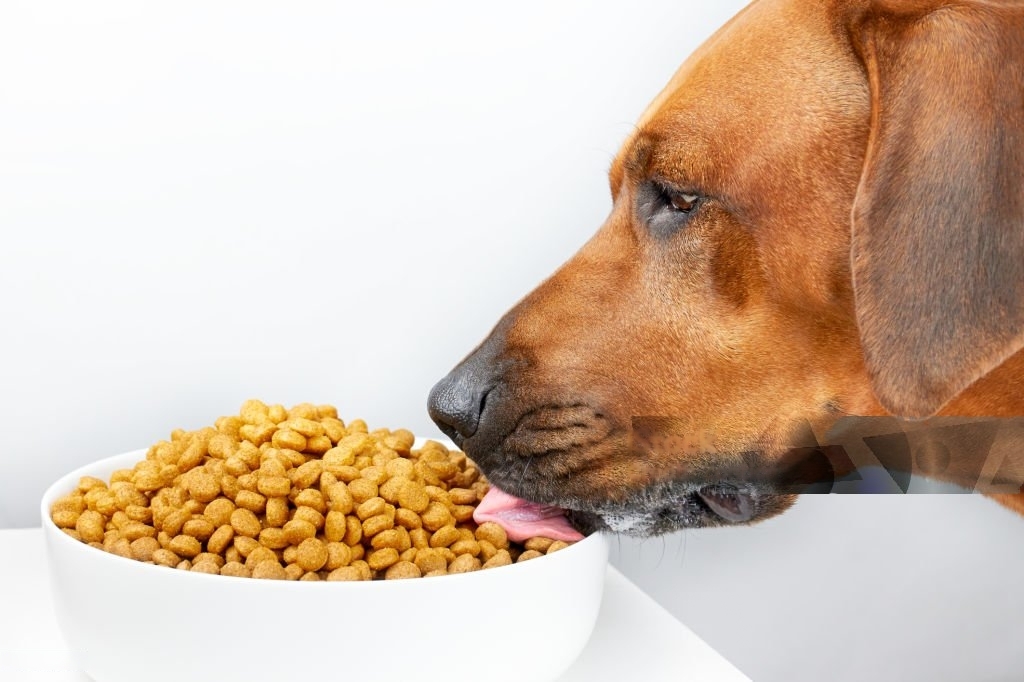
Bloat and Gastric Torsion in Dogs What is BGT? Bloat, or gastric dilatation, and torsion, also known as gastric dilatation-volvulus (GDV), are severe and often life-threatening conditions affecting dogs. These conditions involve the stomach filling with gas and potentially twisting on itself, which can cut off blood supply to vital organs and lead to shock and death if not treated promptly. Understanding the causes, recognizing the symptoms, and implementing preventive measures, including the surgical procedure known as gastropexy, are crucial for dog owners. Understanding Bloat and Gastric Torsion Bloat occurs when a dog’s stomach fills with gas, fluid, or food, causing it to expand. This distension can lead to increased pressure on other organs and the diaphragm, making it difficult for the dog to breathe. When bloat progresses to GDV, the stomach twists around its short axis (volvulus), which traps the gas and prevents the stomach from emptying its contents. This twist also cuts off blood flow to the stomach and spleen, leading to tissue death and a systemic inflammatory response. GDV is a medical emergency that requires immediate veterinary attention. Without prompt treatment, GDV can result in the death of the affected dog within hours. Risk Factors Certain factors can increase the likelihood of a dog developing bloat and GDV, including: Breed and Size: Large and giant breeds with deep chests, such as Great Danes, German Shepherds, and Standard Poodles, are more prone to GDV. Age: Older dogs are at higher risk, although bloat can occur at any age. Genetics: A familial history of bloat increases the risk. Eating Habits: Dogs that eat rapidly, consume large meals, or drink excessive amounts of water before or after meals are more susceptible. Stress: Stressful situations, such as boarding, traveling, or new environments, can precipitate bloat. Symptoms and Diagnosis Early recognition of bloat and GDV symptoms is critical for survival. Symptoms may include: A distended abdomen Unsuccessful attempts to vomit Excessive drooling Restlessness and pacing Rapid, shallow breathing Signs of pain when touched around the abdomen If GDV is suspected, immediate veterinary intervention is necessary. Diagnosis typically involves a physical examination, radiographs (X-rays) to confirm stomach distension and twisting, and possibly blood tests to assess the extent of the condition and organ function. Emergency Treatment Emergency treatment for GDV involves stabilizing the dog and preparing for surgery. Initial steps include: Decompression: Removing the gas from the stomach using a stomach tube or needle to relieve pressure. Intravenous Fluids: Administering fluids to combat shock and support blood pressure. Pain Relief and Antibiotics: Providing medications to manage pain and prevent infection. Surgery is required to untwist the stomach and assess any damage to the stomach and spleen. In severe cases, part of the stomach may need to be removed if the tissue is necrotic. The surgeon may also perform a gastropexy during this procedure to prevent recurrence. Prevention Strategies Preventing bloat and GDV involves several management strategies: Diet and Feeding: Feed smaller, more frequent meals rather than one or two large meals. Use slow-feeder bowls to reduce the speed of eating. Avoid vigorous exercise immediately before and after meals. Provide a calm environment during feeding times to reduce stress. Hydration: Ensure the dog has access to water at all times, but limit excessive drinking immediately after meals. Encourage hydration throughout the day in smaller amounts. Routine Veterinary Care: Regular veterinary check-ups can help monitor the dog’s overall health and catch any early signs of digestive issues. Gastropexy as a Preventive Treatment Gastropexy is a surgical procedure that involves attaching the stomach to the abdominal wall to prevent it from twisting. This procedure can be performed prophylactically (preventively) or during emergency surgery for GDV. There are several techniques for gastropexy, including: Traditional (Open) Gastropexy: Performed through a larger abdominal incision, often during spaying or neutering. Laparoscopic Gastropexy: A minimally invasive approach using small incisions and a camera to guide the surgery. Endoscopic-Assisted Gastropexy: Combines endoscopy and surgery for a less invasive procedure. Benefits and Risks of Gastropexy The primary benefit of gastropexy is the significant reduction in the risk of GDV recurrence. For dogs at high risk, such as those with a familial history or breeds predisposed to GDV, gastropexy can be a lifesaving preventive measure. Conclusion Bloat and gastric torsion are serious conditions that require immediate attention. Understanding the risk factors and symptoms can help dog owners take prompt action to save their pets’ lives. Preventive measures, such as proper feeding practices and stress reduction, are crucial in minimizing the risk. For high-risk dogs, a prophylactic gastropexy can provide significant protection against GDV. By staying informed and vigilant, dog owners can help ensure the health and safety of their canine companions. Any other doubt? We are here to help, Contact us now. Contact Us

Giardiasis
Giardiasis in Dogs: Understanding, Prevention, and Management Giardiasis Giardiasis is a prevalent parasitic infection in dogs caused by the protozoan Giardia intestinalis. This microscopic organism resides in the small intestine, leading to gastrointestinal disturbances that can significantly impact a dog’s health and well-being. Proper deworming practices, recognizing the signs of infection, and understanding the risks associated with giardiasis are crucial for ensuring the health of our canine companions. Understanding Giardiasis Giardia is a protozoan parasite that exists in two forms: the motile trophozoite and the environmentally resistant cyst. Dogs typically become infected by ingesting the cysts, which can be found in contaminated water, food, or feces. Once ingested, the cysts transform into trophozoites in the small intestine, where they attach to the intestinal lining and multiply. Transmission and Risk Factors Giardiasis is highly contagious and can spread rapidly among dogs, especially in environments such as kennels, shelters, and dog parks. The primary mode of transmission is through the fecal-oral route, where dogs ingest contaminated water, food, or come into contact with infected feces. Dogs that groom themselves or other animals can also ingest cysts inadvertently. Several factors can increase the risk of giardiasis in dogs, including: Poor Sanitation: Environments with inadequate hygiene practices are breeding grounds for Giardia cysts. Crowded Living Conditions: Dogs living in close quarters with other dogs are more susceptible to infection. Young Age: Puppies are more prone to giardiasis due to their developing immune systems. Weakened Immune System: Dogs with compromised immune systems are at a higher risk of infection. Access to Contaminated Water: Dogs that drink from puddles, lakes, or streams are at risk of ingesting Giardia cysts. Signs and Symptoms of Giardiasis The symptoms of giardiasis in dogs can vary in severity, ranging from mild to severe gastrointestinal distress. Common signs include: Diarrhea: Often intermittent, foul-smelling, and greasy. Diarrhea may be acute or chronic. Weight Loss: Despite having a normal or increased appetite, infected dogs may lose weight. Lethargy: Affected dogs might appear tired or less active than usual. Vomiting: Some dogs may experience vomiting, although this is less common. Dehydration: Prolonged diarrhea can lead to dehydration, a serious concern that requires immediate attention. Abdominal Discomfort: Dogs may show signs of abdominal pain or discomfort, such as whining or adopting unusual postures. Diagnosing Giardiasis Diagnosing giardiasis can be challenging due to the intermittent shedding of Giardia cysts in feces. Veterinarians typically use a combination of methods to confirm the infection: Fecal Examination: Microscopic examination of fecal samples to detect Giardia cysts or trophozoites. Fecal Antigen Tests: These tests detect specific Giardia antigens in fecal samples and are more reliable than microscopic examination. PCR (Polymerase Chain Reaction): A molecular test that can identify Giardia DNA in fecal samples, providing a definitive diagnosis. Proper Deworming and Prevention Effective deworming and prevention strategies are essential to control giardiasis in dogs. Here are key measures to consider: Regular Deworming: Administering appropriate deworming medications as prescribed by a veterinarian is crucial. Commonly used medications include fenbendazole and metronidazole. Follow the vet’s instructions carefully to ensure complete eradication of the parasite. Hygiene and Sanitation: Maintain clean living conditions for dogs. Regularly clean and disinfect kennels, bedding, and feeding areas to reduce the risk of contamination. Safe Drinking Water: Ensure dogs have access to clean, fresh water. Avoid allowing dogs to drink from potentially contaminated sources such as puddles, lakes, and streams. Prompt Waste Removal: Regularly remove and dispose of feces from yards and living areas to prevent the spread of Giardia cysts. Isolation of Infected Dogs: If a dog is diagnosed with giardiasis, isolate them from other dogs to prevent transmission until the infection is cleared. Managing an Infection If a dog is diagnosed with giardiasis, prompt and effective treatment is necessary to alleviate symptoms and prevent further spread. Treatment typically involves: Medication: Administering prescribed anti-parasitic medications to eliminate the Giardia infection. It is crucial to complete the full course of treatment even if symptoms improve. Supportive Care: Ensuring the dog remains hydrated and providing a balanced diet to support recovery. In severe cases, veterinary intervention may be needed for intravenous fluids and other supportive measures. Follow-Up Testing: Conduct follow-up fecal tests to confirm the infection has been cleared and to prevent reinfection. Conclusion Giardiasis in dogs is a common but manageable condition with appropriate deworming, hygiene practices, and preventive measures. By recognizing the signs and symptoms of giardiasis, maintaining a clean environment, and ensuring regular veterinary care, dog owners can protect their pets from this parasitic infection. Early intervention and diligent management are key to safeguarding the health and happiness of our furry friends. Any other doubt? We are here to help, Contact us now. Contact Us Preventive Medicine Click here Pet Surgery Click here Alternative Medicine Click here Pet Dentistry Click here
