

Mastocytoma Tumors in Dogs Mastocytoma? Mastocytoma tumors, commonly known as mast cell tumors, are a prevalent form of cancer in dogs. These tumors originate from mast cells, which are an essential part of the immune system. While mastocytoma tumors can occur in any breed, they are more commonly found in Boxers, Boston Terriers, Labrador Retrievers, and Golden Retrievers. Understanding the types, signs, and treatment options for mastocytoma tumors is crucial for pet owners and veterinarians alike. Types There are three primary types of mastocytoma tumors in dogs: cutaneous, subcutaneous, and visceral. Cutaneous Mastocytoma Tumors: These tumors develop on the skin and are the most common type seen in dogs. They typically present as firm, raised masses that may vary in size and appearance. Cutaneous mastocytoma tumors are often found on the trunk, limbs, or perineal area of the dog. Subcutaneous Mastocytoma Tumors: These tumors develop beneath the skin, often in the deeper layers of tissue. They may feel like soft lumps under the skin and can be more challenging to detect without thorough examination. Subcutaneous mastocytoma tumors can sometimes be mistaken for lipomas due to their similar texture. Visceral Mastocytoma Tumors: These tumors develop in internal organs such as the spleen, liver, or gastrointestinal tract. Visceral mastocytoma tumors are less common than cutaneous or subcutaneous tumors but can be more challenging to diagnose and treat due to their location. Signs The signs of mastocytoma tumors in dogs can vary depending on the location and severity of the tumor. However, some common signs may include: Visible Masses or Lumps: The presence of firm, raised masses on the skin or beneath the skin. Skin Changes: Redness, swelling, or ulceration of the skin overlying the tumor. Gastrointestinal Symptoms: Vomiting, diarrhea, abdominal pain, or loss of appetite may occur with visceral mastocytoma tumors. Systemic Signs: In advanced cases, dogs may exhibit lethargy, weakness, weight loss, or jaundice. It’s essential for pet owners to monitor their dogs for any unusual lumps or changes in behavior and seek veterinary attention promptly if any concerning signs are noticed. Diagnostic Procedures Diagnosing mastocytoma tumors in dogs typically involves a combination of clinical examination, cytology, imaging studies, and sometimes biopsy. These diagnostic procedures are essential for confirming the presence of a tumor, determining its type and grade, and guiding treatment decisions. Clinical Examination: A thorough physical examination by a veterinarian is the first step in diagnosing mastocytoma tumors. The veterinarian will palpate the dog’s skin and underlying tissues to detect any lumps or masses and assess for signs of inflammation or ulceration. Cytology: Fine-needle aspiration (FNA) or impression smear cytology may be performed to obtain a sample of cells from the suspected tumor. These samples are then examined under a microscope to identify mast cells and evaluate their morphology. Cytology can provide rapid and non-invasive confirmation of a mastocytoma tumor and help determine its grade. Imaging Studies: In cases where there is suspicion of visceral mastocytoma tumors or metastasis, imaging studies such as ultrasound, X-rays, or advanced imaging modalities like computed tomography (CT) or magnetic resonance imaging (MRI) may be performed. These studies can help visualize the extent of the tumor, assess its involvement with surrounding tissues, and identify any spread to other organs. Biopsy: In some cases, particularly if the diagnosis is uncertain or if surgical removal is not immediately feasible, a biopsy may be recommended. A biopsy involves the surgical removal of a tissue sample from the suspected tumor site for histopathological examination. Histopathology allows for a more detailed assessment of the tumor’s characteristics, including its grade, mitotic index, and margins. Blood Tests: Blood tests, including a complete blood count (CBC) and serum biochemistry profile, may be performed to evaluate the dog’s overall health status and assess for any abnormalities that may impact treatment decisions or prognosis. By combining these diagnostic procedures, veterinarians can accurately diagnose mastocytoma tumors, determine their severity, and develop an appropriate treatment plan tailored to the individual needs of the dog. Early diagnosis and intervention are crucial for optimizing treatment outcomes and improving the prognosis for dogs affected by mastocytoma tumors. Regular monitoring and follow-up examinations are essential to monitor treatment response and detect any signs of recurrence or progression. Treatment Options for Mastocytoma Tumors: The treatment approach for mastocytoma tumors in dogs typically involves a combination of surgical excision, chemotherapy, and supportive care. The specific treatment plan will depend on factors such as the tumor’s location, size, grade, and whether it has spread to other parts of the body. Surgical Excision: For localized tumors, surgical removal is often the primary treatment option. The goal is to remove the tumor with wide margins to reduce the risk of recurrence. In some cases, additional surgery may be necessary if the tumor is large or has infiltrated surrounding tissues. Chemotherapy: In cases where the tumor is aggressive or has spread to other parts of the body, chemotherapy may be recommended to help shrink the tumor and slow its progression. Chemotherapy drugs commonly used for mastocytoma tumors include vinblastine, lomustine, and prednisone. Radiation Therapy: In certain cases, radiation therapy may be used either as a primary treatment or in combination with surgery and chemotherapy. Radiation therapy can be particularly beneficial for tumors that are not amenable to surgical removal or have a high risk of recurrence. Supportive Care: Throughout the treatment process, supportive care is essential to help manage symptoms and improve the dog’s quality of life. This may include pain management, nutritional support, and monitoring for any potential side effects of treatment. In conclusion, mastocytoma tumors are a significant health concern for dogs, requiring prompt diagnosis and appropriate treatment. By understanding the types, signs, and treatment options for mastocytoma tumors, pet owners can work closely with their veterinarians to provide the best possible care for their beloved canine companions. Regular veterinary examinations and proactive monitoring are essential for early detection and intervention, ultimately improving the prognosis for dogs affected by mastocytoma tumors. Any other doubt? We are here to help, Contact us now. Contact

Feline Infectious Peritonitis
Feline Infectious Peritonitis (FIP) What is FIP? Feline Infectious Peritonitis (FIP) is a devastating disease that affects cats worldwide. Understanding its impact, contagious nature, and the importance of vaccination is crucial for cat owners and veterinarians alike. FIP is caused by a coronavirus, specifically a mutated form of the feline coronavirus (FCoV). While most cats encounter FCoV without developing FIP, in some cases, the virus mutates and triggers an abnormal immune response, leading to FIP. This disease primarily affects young cats, but cats of any age can be susceptible. The clinical signs of FIP are diverse and often nonspecific, making it challenging to diagnose. Cats with FIP may exhibit symptoms such as fever, weight loss, lethargy, lack of appetite, and a swollen abdomen due to fluid accumulation (known as “wet” FIP) or organ dysfunction (referred to as “dry” FIP). Unfortunately, FIP is usually fatal, and affected cats often succumb to the disease within weeks or months of diagnosis. FIP is not directly contagious between cats. Instead, it is believed that the FCoV, which is shed in the feces and saliva of infected cats, can spread between cats in multi-cat households or densely populated environments. However, it’s important to note that not all cats exposed to FCoV develop FIP. Factors such as the cat’s immune response and genetic predisposition play a role in determining whether FCoV infection progresses to FIP. Preventing FIP is challenging due to the complexities of the disease. However, vaccination is one of the most effective strategies available. The FIP vaccine stimulates the cat’s immune system to produce antibodies against the coronavirus, potentially reducing the severity of FIP if the cat becomes infected. While the vaccine does not provide complete protection, it can help decrease the risk and severity of the disease in vaccinated cats. Vaccination is especially important for cats at higher risk of exposure to FCoV, such as those living in multi-cat households, catteries, or shelters. Additionally, kittens born to queens (mother cats) with a history of FCoV infection may benefit from vaccination to help protect them during their vulnerable early months. However, vaccination decisions should be made in consultation with a veterinarian, considering factors such as the cat’s health status, lifestyle, and potential exposure risks. Additionally, it’s important to note that the FIP vaccine is not recommended for all cats, and its efficacy can vary among individuals. In addition to vaccination, other measures can help reduce the risk of FIP transmission and infection. These include practicing good hygiene, maintaining a clean living environment for cats, minimizing stress, and promptly addressing any health concerns or illnesses in cats to bolster their immune systems. While FIP remains a complex and challenging disease, ongoing research efforts aim to improve our understanding of its causes, transmission dynamics, and potential treatments. By staying informed, working closely with veterinarians, and taking proactive measures such as vaccination, cat owners can help protect their feline companions from the devastating effects of FIP. Any other doubt? We are here to help, Contact us now. Contact Us
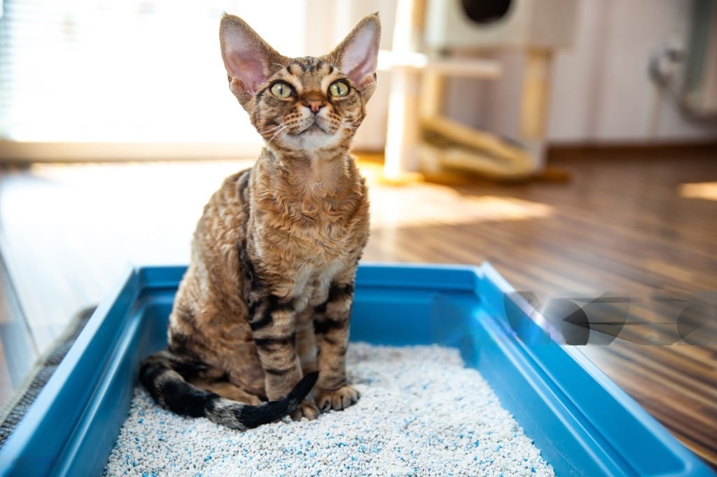
UTO
Understanding Feline Urinary Tract Obstructions What UTO and why it happens? Feline urinary tract obstructions (UTO) are critical emergencies characterized by the partial or complete blockage of the urethra, primarily affecting male cats. This condition impedes the normal flow of urine, leading to severe complications such as kidney damage, electrolyte imbalances, and even death if left untreated. Understanding the causes, clinical signs, treatment options, and breed predispositions associated with feline UTO is crucial for prompt intervention and successful management. Causes of Feline Urinary Tract Obstructions Feline UTO can arise from various factors, including urinary stones (uroliths), urethral plugs, urethral strictures, inflammation, tumors, and congenital abnormalities. Uroliths, comprised of minerals and organic compounds, can obstruct the urethra and predispose cats to recurrent blockages. Urethral plugs, composed of mucus, proteinaceous material, and cellular debris, often occur in association with feline lower urinary tract disease (FLUTD), a multifactorial condition influenced by diet, stress, and environmental factors. Sings Cats with urinary tract obstructions typically exhibit signs of urethral obstruction, including straining to urinate, vocalization, frequent attempts to urinate, and producing small amounts of urine or no urine at all. Affected cats may also display signs of abdominal discomfort, lethargy, anorexia, and vomiting. Prompt recognition of these clinical signs is essential for initiating timely treatment and preventing life-threatening complications. Breed Predispositions While urinary tract obstructions can affect cats of any breed or age, certain breeds appear to be predisposed to developing urolithiasis and FLUTD. For example, Persian cats, with their brachycephalic skull conformation, may be more prone to urinary tract abnormalities due to altered urinary tract dynamics. Additionally, breeds such as Siamese, Burmese, and Himalayan cats have been reported to have a higher incidence of FLUTD, possibly due to genetic predispositions or environmental factors. Diagnostic Evaluation Diagnostic evaluation of cats with suspected urinary tract obstructions typically involves a thorough physical examination, urinalysis, bloodwork, and imaging studies such as radiography and ultrasonography. Urinalysis may reveal abnormalities such as hematuria, pyuria, and crystalluria, indicative of underlying urinary tract pathology. Radiographs and ultrasound can aid in identifying the presence of urinary stones, urethral strictures, or bladder abnormalities contributing to the obstruction. Treatment Options The management of feline urinary tract obstructions aims to relieve urethral obstruction, correct electrolyte imbalances, alleviate pain, and address underlying predisposing factors. Immediate intervention is often required to alleviate urethral obstruction and restore normal urine flow. Treatment modalities may include: Urethral catheterization: Passage of a urinary catheter under sedation or anesthesia to dislodge and remove obstructive material from the urethra. Urinary catheterization: Placement of a urinary catheter to facilitate continuous urine drainage and relieve bladder distention. Intravenous fluid therapy: Administration of intravenous fluids to correct dehydration, restore electrolyte balance, and support renal function. Analgesic therapy: Provision of analgesic medications to alleviate pain and discomfort associated with urinary tract obstruction. Dietary management: Prescription diets formulated to dissolve or prevent the formation of urinary stones and reduce the risk of recurrent obstructions. Long-Term Management and Prevention Following initial stabilization, long-term management of feline urinary tract obstructions focuses on identifying and addressing predisposing factors, implementing dietary modifications, and monitoring for recurrence of clinical signs. Regular veterinary examinations, urinalysis, and imaging studies may be recommended to assess urinary tract health and identify early signs of recurrence or complications. Conclusion Feline urinary tract obstructions represent critical emergencies that require prompt recognition, intervention, and comprehensive management. By understanding the underlying causes, clinical presentation, diagnostic evaluation, treatment options, and breed predispositions associated with feline UTO, veterinarians and pet owners can collaborate to optimize patient outcomes and minimize the risk of recurrence. Early intervention and proactive management strategies are essential for preserving urinary tract health and enhancing the quality of life for affected cats. Any other doubt? We are here to help, Contact us now. Contact Us
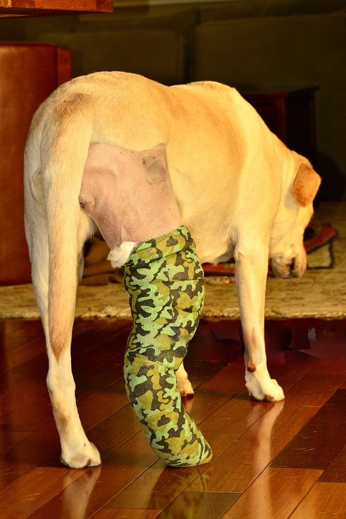
CCLR
Understanding Cranial Cruciate Ligament Rupture in Dogs What is CCL ? Cruciate Ligament (CCL) rupture is one of the most common orthopedic injuries in dogs, affecting their hind limbs. This ligament, analogous to the anterior cruciate ligament (ACL) in humans, plays a crucial role in stabilizing the knee joint. When ruptured, it can lead to lameness, pain, and osteoarthritis. Understanding the dynamics of CCL rupture, along with breed-specific considerations, diagnostics, and treatment options, is pivotal for effective management and rehabilitation in affected dogs. Why does this happen? The CCL in dogs is susceptible to injury due to its role in stabilizing the knee joint during weight-bearing activities and sudden changes in direction. Rupture often occurs due to chronic degenerative changes or acute trauma. Breeds predisposed to CCL rupture, such as Labrador Retrievers, Rottweilers, and Bulldogs, may exhibit varying degrees of ligament laxity due to genetic factors or conformational abnormalities. Sings Dogs with CCL rupture typically present with acute or progressive hind limb lameness, reluctance to bear weight, and difficulty rising or climbing stairs. Physical examination may reveal joint effusion, pain on manipulation, and instability upon cranial drawer or tibial compression tests. Moreover, chronic cases may exhibit signs of osteoarthritis, including joint crepitus and muscle atrophy. Diagnostics Diagnostic imaging, such as radiography and advanced techniques like magnetic resonance imaging (MRI) or computed tomography (CT), plays a crucial role in confirming CCL rupture and assessing concurrent joint pathology. Radiographs may reveal signs of osteoarthritis, joint effusion, and cranial tibial thrust. MRI and CT scans offer detailed visualization of ligament integrity, meniscal injury, and cartilage degeneration, aiding in treatment planning and prognostication. Treatment Options Treatment of CCL rupture in dogs varies based on the severity of clinical signs, concurrent joint pathology, and owner preferences. Conservative management, including rest, anti-inflammatory medications, physical therapy, and weight management, may be suitable for small breed dogs or cases with mild clinical signs and minimal joint instability. However, surgical intervention remains the gold standard for restoring joint stability and preventing progressive osteoarthritis. Surgical Techniques Various surgical techniques have been developed to address CCL rupture in dogs, including extracapsular stabilization, tibial plateau leveling osteotomy (TPLO), and tibial tuberosity advancement (TTA). Extracapsular techniques, such as the lateral fabellar suture, involve stabilizing the joint using synthetic materials outside the joint capsule. TPLO and TTA techniques alter the biomechanics of the knee joint by changing the angle of the tibial plateau, thereby reducing cranial tibial thrust and restoring joint stability. Breed-Specific Considerations Certain dog breeds exhibit unique anatomical and biomechanical characteristics that may influence the development and management of CCL rupture. For instance, brachycephalic breeds like Bulldogs may have altered joint angles and increased risk of ligament injury due to their conformational traits. Additionally, large and giant breeds may experience accelerated progression of osteoarthritis following CCL rupture, necessitating early intervention and comprehensive rehabilitation strategies. Conclusion Cranial Cruciate Ligament rupture is a prevalent orthopedic condition in dogs, with significant implications for mobility and quality of life. Prompt recognition, accurate diagnosis, and appropriate treatment are essential for minimizing pain, preventing joint degeneration, and optimizing long-term outcomes. By understanding the pathophysiology, diagnostics, treatment options, and breed-specific considerations associated with CCL rupture, veterinarians and pet owners can collaborate to provide comprehensive care tailored to the individual needs of affected dogs. Any other doubt? We are here to help, Contact us now. Contact Us
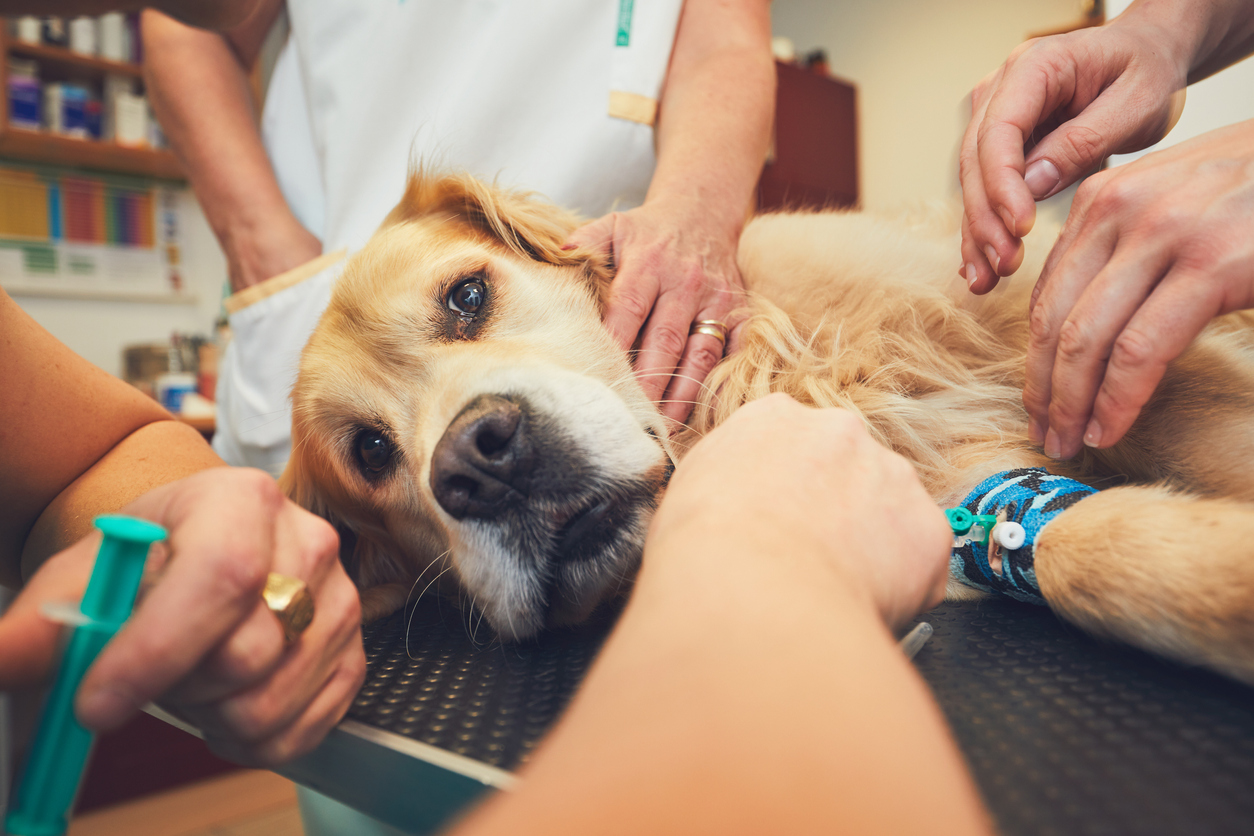
Parvo
Parvovirus in Dogs and the Vital Role of Vaccination Protocols What is Parvovirosis? Parvovirus, a highly contagious and potentially fatal viral disease, poses a significant threat to dogs worldwide. Understanding the nature of parvovirus, its symptoms, treatment options, and most importantly, the role of vaccination protocols, is crucial for dog owners and veterinarians alike in safeguarding canine health and well-being. Parvovirus: The Disease and Its Impact Parvovirus, caused by canine parvovirus type 2 (CPV-2), primarily affects the gastrointestinal tract and immune system of dogs. The virus is highly resilient and can survive in the environment for extended periods, making it easily transmissible through contact with contaminated feces, surfaces, or objects. The symptoms of parvovirus infection often include severe vomiting, diarrhea (often bloody), lethargy, loss of appetite, and dehydration. Puppies and unvaccinated dogs are particularly susceptible to parvovirus, and the disease can progress rapidly, leading to life-threatening complications such as septicemia and organ failure if left untreated. Treatment Challenges and Prognosis Treating parvovirus can be challenging and intensive, often requiring hospitalization, intravenous fluid therapy, antibiotics to prevent secondary infections, antiemetics to control vomiting, and supportive care to maintain hydration and nutrition. Despite aggressive treatment, the prognosis for dogs with severe parvovirus infection remains guarded, with mortality rates ranging from 10% to 30% or higher in some cases. Early detection and prompt intervention are critical in improving the chances of survival and reducing the severity of complications associated with parvovirus infection. However, prevention through vaccination remains the most effective strategy in protecting dogs against this devastating disease. The Importance of Vaccination Protocols Vaccination is a cornerstone of preventive healthcare for dogs and plays a central role in reducing the risk of parvovirus infection. Puppies are particularly vulnerable to parvovirus due to their immature immune systems, making timely vaccination essential in providing early protection. A proper vaccination protocol typically involves a series of initial vaccinations administered to puppies starting at around 6 to 8 weeks of age, with booster doses given every 3 to 4 weeks until the puppy reaches 16 weeks of age or older. Adult dogs require regular booster vaccinations to maintain immunity against parvovirus and other preventable diseases. Vaccines stimulate the dog’s immune system to produce protective antibodies against parvovirus, thereby priming the body’s defenses to recognize and neutralize the virus upon exposure. Vaccination not only helps prevent clinical disease but also reduces the risk of virus shedding, thereby contributing to community-wide disease control efforts. Challenges and Considerations While vaccination offers significant protection against parvovirus, several factors can impact vaccine efficacy, including improper storage and handling of vaccines, maternal antibody interference in young puppies, and variations in individual immune response. It is essential for dog owners to work closely with their veterinarians to develop a tailored vaccination schedule based on the dog’s age, health status, lifestyle, and risk factors. Regular veterinary examinations provide an opportunity to assess the dog’s vaccination status, discuss preventive healthcare measures, and address any concerns or questions regarding vaccination protocols. In conclusion, parvovirus remains a serious threat to the health and well-being of dogs worldwide, highlighting the importance of comprehensive vaccination protocols in preventing the spread of this devastating disease. By adhering to recommended vaccination schedules and partnering with veterinarians to implement preventive healthcare strategies, dog owners can help ensure their canine companions lead healthy, happy lives free from the scourge of parvovirus infection. Any other doubt? We are here to help, Contact us now. Contact Us

Hyperthyroid cat
Hyperthyroidism in Cats: Understanding, Treatment, and the Importance of Diligent Veterinary Follow-ups What is Hyperthyroidism? Hyperthyroidism is a common endocrine disorder affecting cats, particularly older felines. Characterized by an overactive thyroid gland, this condition can lead to a variety of health issues if left untreated. Understanding the pathology, treatment options, and the significance of regular veterinary follow-ups is crucial in managing hyperthyroidism in cats effectively. Pathology of Hyperthyroidism: The thyroid gland, located in a cat’s neck, produces hormones that regulate metabolism. In hyperthyroidism, the thyroid gland becomes overactive, producing excessive amounts of thyroid hormones, primarily thyroxine (T4). This overproduction disrupts the body’s metabolic balance, leading to a range of symptoms. The exact cause of hyperthyroidism in cats remains unclear, although several factors, including genetic predisposition, diet, and environmental influences, may contribute to its development. Additionally, benign tumors known as thyroid adenomas often form within the thyroid gland, further exacerbating hormone production. Symptoms and Diagnosis: Hyperthyroidism manifests through various symptoms, including weight loss despite an increased appetite, hyperactivity, excessive thirst and urination, vomiting, diarrhea, and poor coat quality. Cats may also exhibit behavioral changes, such as restlessness and aggression. Diagnosing hyperthyroidism typically involves a thorough physical examination, blood tests to measure thyroid hormone levels (T4), and sometimes additional imaging tests like ultrasound to evaluate the thyroid gland’s size and structure. Treatment Options: Several treatment options are available to manage hyperthyroidism in cats, each with its benefits and considerations: Medication: Anti-thyroid drugs such as methimazole work by inhibiting the production of thyroid hormones. While effective, long-term medication administration may be necessary, and regular monitoring of thyroid levels is essential to adjust dosage as needed. Radioactive Iodine Therapy: Considered the gold standard treatment for hyperthyroidism, radioactive iodine therapy involves a single injection of radioactive iodine, which selectively targets and destroys overactive thyroid tissue. This treatment offers a high success rate and potential cure for hyperthyroidism but requires specialized facilities due to radiation safety protocols. Surgical Thyroidectomy: Surgical removal of the thyroid gland is another treatment option, particularly for cats who are not candidates for radioactive iodine therapy. While effective, surgery carries risks, including anesthesia complications and potential damage to nearby structures. Dietary Management: Prescription diets formulated with limited iodine content can help manage hyperthyroidism by reducing thyroid hormone production. While not curative, dietary management may complement other treatment modalities or serve as a primary therapy for cats with mild hyperthyroidism. Importance of Diligent Follow-ups: Regular veterinary follow-ups are essential in managing hyperthyroidism and ensuring the long-term health and well-being of affected cats. These follow-ups serve several critical purposes: Monitoring Response to Treatment: Veterinary check-ups allow veterinarians to assess the cat’s response to treatment, monitor thyroid hormone levels, and adjust medication dosages as needed. Regular blood tests help ensure that thyroid hormone levels remain within the optimal range, minimizing the risk of under- or over-treatment. Detecting Complications: Hyperthyroidism can predispose cats to various complications, including heart disease, hypertension, and kidney dysfunction. Routine examinations enable early detection of these complications, facilitating prompt intervention and management. Addressing Emerging Health Concerns: As cats age, they may develop concurrent health issues unrelated to hyperthyroidism. Regular veterinary visits provide an opportunity to address emerging health concerns, administer preventive care, and discuss strategies for optimizing the cat’s quality of life. In conclusion, hyperthyroidism is a common endocrine disorder affecting older cats, characterized by excessive thyroid hormone production. Treatment options include medication, radioactive iodine therapy, surgical thyroidectomy, and dietary management. Diligent veterinary follow-ups are essential for monitoring response to treatment, detecting complications, and addressing emerging health concerns. By working closely with veterinarians and adhering to recommended follow-up schedules, cat owners can effectively manage hyperthyroidism and improve their feline companions’ overall health and well-being. Any other doubt? We are here to help, Contact us now. Contact Us
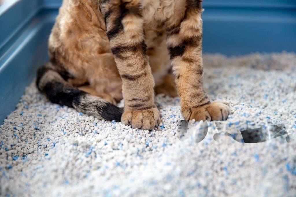
Urolithiasis in cats
Managing Urolithiasis in Cats: The Role of Prescription Diets in Treatment What is Urolithiasis? Urolithiasis, a condition characterized by the formation of urinary stones, can pose significant health risks to our feline companions. Understanding the causes, symptoms, and treatment options, particularly the role of prescription diets, is crucial in effectively managing urolithiasis and promoting the well-being of cats. Urolithiasis occurs when minerals in the urine crystallize and form stones within the urinary tract. While the exact cause of urolithiasis in cats can vary, factors such as diet, water intake, genetics, and underlying medical conditions may contribute to stone formation. Common types of urinary stones in cats include struvite, calcium oxalate, and urate stones. Recognizing the signs of urolithiasis is essential for early intervention. Symptoms may include: Straining to urinate Frequent urination Blood in the urine Urinating outside the litter box Vocalization or signs of discomfort during urination Lethargy and decreased appetite If you notice any of these symptoms in your cat, it’s crucial to seek veterinary care promptly. Delayed treatment can lead to complications such as urinary tract obstruction, kidney damage, and systemic infection, which can be life-threatening. Upon examination, veterinarians may perform diagnostic tests such as urinalysis, urine culture, blood tests, and imaging studies (e.g., X-rays, ultrasound) to confirm the diagnosis and identify the type and location of urinary stones. Treatment options for urolithiasis in cats may include medical management, dietary modification, and, in some cases, surgical intervention. Prescription diets play a vital role in the management of urolithiasis, as they are specifically formulated to dissolve or prevent the formation of certain types of urinary stones. For cats with struvite urolithiasis, prescription diets formulated to promote urinary acidification and reduce the concentration of magnesium, phosphorus, and protein can help dissolve existing stones and prevent recurrence. These diets typically contain controlled levels of minerals and ingredients that promote urine dilution and maintain a healthy urinary pH. On the other hand, cats with calcium oxalate or urate urolithiasis require diets that promote urinary dilution and discourage the formation of crystals and stones. These diets may have reduced levels of calcium, oxalate, and purines, along with increased moisture content to encourage water intake and urinary flushing. It’s essential to follow your veterinarian’s recommendations regarding the selection and use of prescription diets for managing urolithiasis in cats. Feeding guidelines, feeding schedules, and monitoring protocols should be strictly adhered to in order to optimize the effectiveness of the prescribed diet and minimize the risk of complications. In addition to prescription diets, other management strategies for urolithiasis may include: Encouraging increased water intake through wet food, water fountains, or multiple water sources Providing access to clean litter boxes and ensuring proper litter box hygiene Monitoring urinary habits and behaviors for any changes or abnormalities Administering medications as prescribed by your veterinarian to manage pain, inflammation, or urinary tract infections Regular veterinary examinations and monitoring are essential for cats diagnosed with urolithiasis to assess their response to treatment, monitor for recurrence, and address any underlying health concerns. In conclusion, urolithiasis is a common and potentially serious condition in cats that requires comprehensive management and veterinary care. Prescription diets play a crucial role in the treatment of urolithiasis by promoting urinary health, dissolving stones, and preventing recurrence. By working closely with your veterinarian and following their recommendations, you can help improve your cat’s quality of life and reduce the risk of urinary stone formation and associated complications. Any other doubt? We are here to help, Contact us now. Contact Us
Pyometra
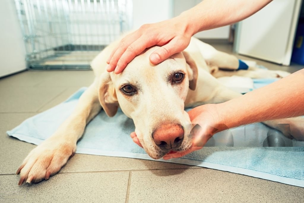
Understanding Pyometra: A Common Yet Serious Condition in Female Dogs h What is pyometra? Pyometra, though it might sound intimidating, is a condition that commonly affects unspayed female dogs. It’s essential to grasp the basics of this condition, its symptoms, treatment, and prevention, as it can significantly impact your furry friend’s health and well-being. Firstly, let’s break down the term “pyometra.” “Pyo” refers to pus, and “metra” pertains to the uterus. Essentially, pyometra translates to a pus-filled uterus, which arises due to hormonal changes associated with the reproductive cycle of female dogs. During a dog’s heat cycle, her uterus undergoes changes to prepare for potential pregnancy. If pregnancy doesn’t occur, the uterus remains receptive to bacterial invasion. Bacteria can ascend through the cervix into the uterus, leading to infection and accumulation of pus. This accumulation creates a dangerous environment within the uterus, potentially causing severe illness or even death if left untreated. Recognizing the symptoms of pyometra is crucial for early intervention. While symptoms may vary, common signs include: Increased drinking and urination Loss of appetite Lethargy or weakness Vaginal discharge, which may be pus-like or bloody Abdominal distension or swelling If you notice any of these signs in your furry companion, prompt veterinary attention is imperative. Delayed treatment can escalate the severity of the condition, making it more challenging to manage. When diagnosing pyometra, veterinarians typically conduct a thorough physical examination, along with blood tests, ultrasound, or radiographs to confirm the diagnosis. Once diagnosed, treatment options may include: Emergency surgery to remove the infected uterus (ovariohysterectomy or spaying) Intravenous fluids and antibiotics to stabilize the dog’s condition and fight the infection Surgery is the treatment of choice for pyometra, as it effectively removes the infected uterus and prevents recurrence. While surgery carries inherent risks, the benefits of removing the infected organ far outweigh the risks associated with untreated pyometra. Prevention is key in managing pyometra and safeguarding your dog’s health. Spaying your female dog before her first heat cycle significantly reduces her risk of developing pyometra. Spaying eliminates the hormonal fluctuations that predispose the uterus to infection, offering long-term health benefits for your pet. It’s essential to address common misconceptions surrounding spaying and neutering. Some pet owners worry that spaying their female dogs will alter their personality or cause weight gain. However, the benefits of spaying, including preventing pyometra, outweigh these concerns. Moreover, proper diet and exercise can help maintain your dog’s weight and overall well-being. In conclusion, pyometra is a serious condition that requires prompt veterinary attention. Understanding its causes, symptoms, treatment, and prevention measures empowers pet owners to advocate for their furry companions’ health. By recognizing the signs of pyometra and prioritizing preventive care, we can ensure our beloved dogs live long, healthy lives free from the threat of this potentially life-threatening condition. Any other doubt? We are here to help, Contact us now. Contact Us

External Parasite control
External Parasite Control External parasite control? External parasite control, particularly flea and tick management, is crucial for keeping our furry friends happy and healthy. Dogs, being playful and explorative creatures, often find themselves exposed to these pesky critters while frolicking outdoors. But fear not! With the right protocol in place, we can help protect our canine companions from the discomfort and potential health risks associated with fleas and ticks. First things first, let’s understand the enemy: fleas and ticks. Fleas are tiny, wingless insects that survive by feeding on the blood of animals, including dogs. These little jumpers can cause itching, irritation, and even allergic reactions in our pups. On the other hand, ticks are arachnids that latch onto their hosts, including dogs, to feed on blood. They can transmit diseases such as Lyme disease and Rocky Mountain spotted fever, posing serious health threats. Now, onto the battle plan: the protocol for external parasite control in dogs. Regular Inspection: The first line of defense is vigilance. Make it a habit to inspect your dog regularly for any signs of fleas and ticks. Run your fingers through their fur, paying close attention to areas like the neck, ears, belly, and tail, where these parasites often hide. Preventive Products: Prevention is key! Consult with your veterinarian to determine the most suitable preventive products for your dog. These may include topical treatments, oral medications, or collars designed to repel and kill fleas and ticks. Ask your Vet about the different products specifically formulated for dogs and the best treatment for your furry friend, as those for other animals may contain ingredients that can be harmful to them. Consistent Treatment: Consistency is crucial when it comes to parasite control. Follow the recommended dosage and application instructions provided by your veterinarian or the product manufacturer. Some treatments require monthly application, while others may offer longer-lasting protection. Stick to the schedule to ensure continuous defense against fleas and ticks. Environmental Management: Fleas and ticks don’t just lurk on your dog; they can also infest your home and yard. Vacuum your home regularly, paying special attention to areas where your dog spends time. Wash your dog’s bedding and toys frequently in hot water to kill any parasites and their eggs. Consider using environmental control products, such as sprays and powders, to target fleas and ticks in your living spaces. Year-Round Protection: Don’t let your guard down, even during the colder months. While fleas and ticks may be less active in winter, they can still pose a threat, especially in regions with milder climates. Maintain year-round protection to keep your dog safe from infestations and disease transmission. Monitoring and Adaptation: Keep an eye on your dog for any signs of adverse reactions to preventive products. Some dogs may be sensitive to certain ingredients, experiencing skin irritation or other symptoms. If you notice any unusual reactions, consult your veterinarian promptly. They can recommend alternative products or adjust the treatment protocol as needed. Regular Check-ups: Schedule regular check-ups with your veterinarian to assess your dog’s overall health and discuss their parasite control regimen. Your vet can provide valuable guidance and make adjustments based on your dog’s specific needs and any changes in their environment or lifestyle. By implementing a comprehensive protocol for external parasite control, you can help safeguard your dog against the nuisance and dangers posed by fleas and ticks. With proper care and attention, you can keep your canine companion happy, healthy, and itch-free for years to come! Any other doubt? We are here to help, Contact us now. Contact Us
Cat vaccines
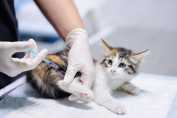
Cat Vaccination Protocols: A Comprehensive Guide When and which vaccines are important for my cat. Ensuring the health and well-being of our feline companions is paramount, and vaccination plays a crucial role in preventing various infectious diseases in cats. A well-structured vaccination protocol is essential to provide optimal protection for our furry friends. In this guide, we will delve into the key aspects of cat vaccination, including the importance of vaccinations, common vaccines, and the recommended vaccination schedule. Importance of Cat Vaccinations: Vaccinations are a cornerstone of preventive healthcare for cats, safeguarding them against potentially life-threatening diseases. These vaccinations stimulate the immune system to produce protective antibodies, preparing the cat’s body to defend itself against specific pathogens. By vaccinating cats, we not only protect individual animals but also contribute to the overall health and well-being of the feline population. Common Cat Vaccines: Feline Viral Rhinotracheitis (FVR) and Feline Calicivirus (FCV): These vaccines target two highly contagious respiratory viruses that can cause severe respiratory symptoms in cats. FVR and FCV vaccinations are typically combined into a single vaccine. Feline Panleukopenia (FP): Also known as feline distemper, FP is a highly contagious and potentially fatal viral disease. Vaccination against panleukopenia is a core component of a cat’s vaccination protocol. Feline Leukemia Virus (FeLV): FeLV is a common viral infection that can lead to immune suppression, anemia, and lymphoma in cats. Vaccination is recommended, especially for cats at higher risk of exposure, such as those that roam outdoors. Rabies: Rabies is a deadly zoonotic disease that can affect both cats and humans. Vaccination against rabies is often mandated by local regulations and is a crucial part of responsible pet ownership. Vaccination Schedule: The vaccination schedule for cats is typically divided into two phases: kittenhood and adulthood. Kitten Vaccination Schedule: Kittens usually receive their first vaccinations at around 6-8 weeks of age. The initial series of vaccines includes FVR, FCV, FP, and FeLV. Boosters are administered every 3-4 weeks until the kitten is around 16 weeks old. Adult Cat Vaccination Schedule: After completing the initial kitten vaccinations, adult cats require regular booster shots to maintain immunity. Booster schedules may vary based on the specific vaccines used and the cat’s lifestyle, but they are generally recommended annually or as advised by the veterinarian. Considerations for Vaccination: Lifestyle Factors: The cat’s lifestyle, such as whether it lives indoors or outdoors, plays a significant role in determining the appropriate vaccination protocol. Outdoor cats, for example, may need additional vaccinations like FeLV. Health Status: Cats with underlying health conditions may have special vaccination requirements. It is crucial for owners to communicate any health concerns to the veterinarian to tailor the vaccination plan accordingly. Local Regulations: Compliance with local regulations regarding specific vaccinations, especially rabies, is essential. Failure to adhere to these regulations may lead to legal consequences and put the cat and other animals at risk. Conclusion: A well-designed cat vaccination protocol is essential for maintaining the health and longevity of our feline companions. By understanding the importance of vaccinations, being aware of common vaccines, and following an appropriate vaccination schedule, cat owners can contribute to a healthier and safer environment for their beloved pets. Regular veterinary consultations are crucial to assess the individual needs of each cat and adjust the vaccination plan accordingly. Remember, a proactive approach to feline health through vaccinations is a key element in providing comprehensive care for our cherished feline friends. Any other doubt? We are here to help, Contact us now. Contact Us
